Monoclonal antibodies represent the rapidly expanding class of biotherapeutics having applications in the treatment of cancer, autoimmune disorders, metabolic disorders, and infectious diseases. One of the major challenges in mAb-based therapeutics development lies in the significant issue of aggregation which in turn has an impact on the quality, safety, and efficacy of the antibody drug. The presence of aggregates in the drug substance or final drug product potentially affects its biological activity thereby making it a critical quality attribute to be assessed. State-of-the-art analytical techniques are required for quality control and detailed characterization. This review provides insight into monoclonal antibody aggregation and the analytical techniques used to detect and quantify these aggregates.
Introduction
Monoclonal antibody (mAb) based therapeutics are assuming a progressively crucial role in the treatment of several ailments such as cancer, autoimmune disorders, and infectious diseases. However, the development of mAbs into well-defined antibody therapeutics that conform to regulatory expectations presents significant challenges due to the complexities of multi-domain mAbs in terms of their physiochemical attributes, including aggregation, fragmentation, charge variations, identity, carbohydrate composition, and higher-order structure (Laptoš & Omersel, 2018).
The aforementioned attributes are tested at various stages of in-process sampling, lot release, stability testing, and analytical comparability assessment in research and development and manufacturing divisions of biopharmaceutical enterprises to gain insights into their structural, conformational, and stability features. This characterization is indispensable for ensuring the quality, safety, and effectiveness of mAbs, particularly in the context of pharmaceutical development and manufacturing (Kaur, 2021).
A spectrum of analytical methods is developed to evaluate the physicochemical attributes of the monoclonal antibody drug to thoroughly investigate the identity, heterogeneity, impurity, and activity of each new batch of monoclonal antibodies (mAbs). This is achieved by techniques such as Size-exclusion chromatography (SEC), Dynamic Light Scattering (DLS), and Analytical Ultracentrifugation (AUC) to determine the molecular weight and size distribution of the mAb, primary structure can be evaluated using advanced methodologies like mass spectrometry and peptide mapping. Additionally, the higher-order structure of the mAb demands scrutiny, encompassing the analysis of secondary, tertiary, and quaternary structures to ensure correct folding and assembly. To achieve this, techniques such as circular dichroism, Fourier-transform infrared spectroscopy, and nuclear magnetic resonance are applied. Furthermore, the identification and quantification of diverse charge variants arising from post-translational modifications, including deamidation, oxidation, and glycosylation are pivotal, requiring the use of capillary electrophoresis and ion-exchange chromatography. The glycosylation pattern attached to the mAb's Fc region is examined in detail through techniques like LC (liquid chromatography) coupled with mass spectrometry (LC-MS). Evaluating the mAb's stability under varying temperature conditions is crucial for which techniques like differential scanning calorimetry and thermal denaturation studies are employed. Furthermore, the impact of osmolality and pH on mAb stability and aggregation propensity is evaluated. Binding affinity, a key parameter, is measured through techniques like surface plasmon resonance or enzyme-linked immunosorbent assays (Le Basle et al., 2020). Spectroscopic methods, including UV-visible spectroscopy and fluorescence spectroscopy, are harnessed to explore the mAb's absorbance, emission, and fluorescence properties. Furthermore, the detection and characterization of sub-visible particles in the mAb solution is performed using techniques such as light obscuration, microscopy, and flow imaging. Also, the solubility profile and aggregation kinetics of mAbs are rigorously evaluated under various conditions to comprehensively understand their physicochemical properties thereby aiding researchers and manufacturers to ensure the quality, stability, and efficacy of mAbs throughout their development, production, and shelf life (Maiti et al., 2023).
This review primarily focuses on mAb aggregation and various methods for characterizing aggregates, a key aspect of physical instability.
Aggregation Insights
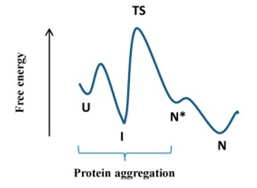
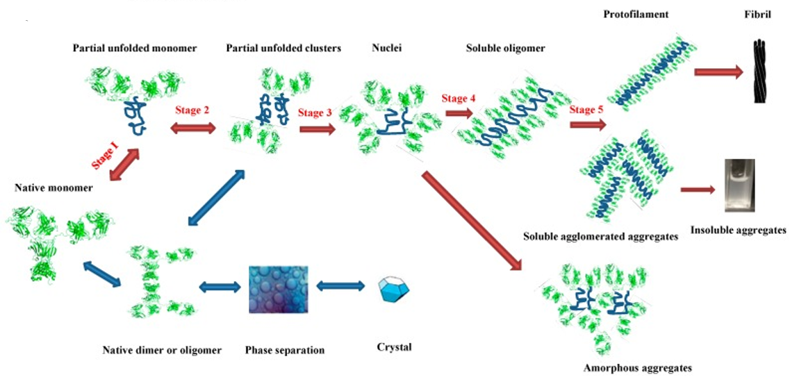
mAb aggregation transforms native proteins from folded states into multimers which are high molecular weight species, irrespective of their size or the nature of bonds connecting them. Soluble or soluble aggregates may arise solely from weak nonspecific interactions or involve covalent bonds like disulfide bonds. In most cases, irreversible Aggregation may occur, especially at advanced stages, and frequently involves proteins adopting non-native conformations. The process begins with a conformational change which is reversible to a higher free energy transition state, which may revert to the native state or form more aggregation-prone intermediates due to decreased free energy after each new aggregate is formed as shown in Figure 1. Protein unfolding exposes hydrophobic residues, that are typically concealed in the native conformation, reducing solubility in hydrophilic buffers and promoting self-association and subsequent aggregation (van der Kant et al., 2017).
|
|
|
a) |
|
|
|
b) |
|
Figure 1. Protein folding process and aggregation (Li et al., 2016) |
Aggregation is associated with heightened immunogenicity of protein therapeutics in patients, potentially triggering immune responses. The degree of immunogenicity appears to increase with aggregate size but is also influenced by glycosylation, product origin, and the presence of contaminants. Immunogenicity can lead to the neutralization of the mAb, resulting in diminished efficacy, or cross-reactions with endogenous counterparts, leading to IgE-mediated immediate hypersensitivity and anaphylaxis.
Under certain conditions, aggregates may precipitate, forming insoluble particles, consisting of proteins or being heterogeneous with excipients and contaminants. Injectable drug solutions, including mAbs, should be free of visible particles, with established limits for sub-visible particles exceeding 10 μm and 25 μm. Enhancing the intrinsic properties of antibodies may result in improved manufacturability, attrition rates, safety, formulation, titers, immunogenicity, and solubility.
Analyzing protein aggregation is challenging due to its wide size range spanning from a few nanometers to a few millimeters. Therefore, a combination of different techniques is used to detect these aggregates (São Pedro et al., 2022). To reduce the likelihood of protein therapeutic aggregates, it is essential to use complementary techniques to analyze aggregates, as outlined in the United States Pharmacopeia (USP) chapter <1787>. Various characterization techniques, such as SEC, UV, DLS, and AUC are explored to predict and mitigate the propensity for monoclonal antibody aggregation (Ilochonwu et al., 2020).
Analytical Techniques for Aggregate Detection
SEC (Size Exclusion Chromatography)
High-performance size-exclusion chromatography (SEC) is the most commonly used technique to estimate the molecular mass of biomolecules thus making it particularly useful for analyzing mAb aggregates in diverse bioprocess stages (São Pedro et al., 2022). Its ease of use, ability to measure low levels of aggregates with minimal sample, short and well-defined separation times are some of the positive attributes that make SEC the method of choice for researchers (Wang et al., 2024).
During the upstream process of mAb production, various components are released by cells into the culture media such as cell debris, host cell proteins, etc. To study mAb aggregates, Affinity chromatography is often used before SEC to selectively purify mAbs and remove these interfering components (Saldanha et al., 2023).
However, there is a caveat when using this two-step approach. The purification step itself can sometimes lead to the formation of aggregates causing misrepresentation of the aggregate content present in the cell culture. To address this concern, a protocol has been developed by Paul et al. for aggregate quantification using SEC without the need for a pre-purification step providing a more direct and accurate assessment of the actual aggregate content in the cell culture (Paul et al., 2017).
Furthermore, the choice of the SEC column is crucial in this process. Traditional SEC columns like TSKgel G3000SWXL may not provide sufficient separation of mAb monomers from other signals in the supernatant thereby complicating the aggregate detection. This is particularly important because accurate quantification of aggregates relies on distinguishing them from other components in the sample. Specialized columns such as MAbPac SEC-1, specifically designed for mAb analysis, offer higher separation efficiencies, and improved separation facilitating accurate quantification of aggregates (Sadighi et al., 2024).
In summary, SEC in combination with appropriate sample preparation techniques and specialized columns is a valuable approach to assessing mAb aggregates, contributing to the development of high-quality and safe biopharmaceutical products.
Dynamic Light Scattering (DLS)
Dynamic Light Scattering (DLS) works by measuring the Brownian motion of particles suspended in a liquid medium to determine their hydrodynamic radius. This motion is influenced by the size of the particles (Sharma et al., 2023). Some of the crucial advantages offered by DLS that make it an attractive choice for researchers and scientists are minimal sample preparation, quick and straightforward measurements, and suitability for high-throughput analysis.
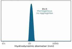
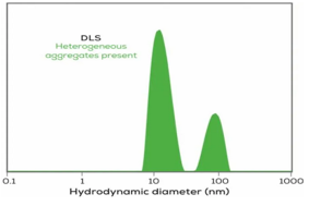
Despite its advantages, DLS has some limitations in detecting low levels of aggregates and in analyzing highly concentrated solutions. In such cases, the technique may struggle to provide accurate measurements due to the weaker scattering signals from small or diluted particles (Bansal et al., 2019). A noteworthy feature of DLS is that its sensitivity increases with particle size. Larger particles scatter light more intensely, making it particularly effective in detecting even trace amounts of larger aggregate molecules as shown in Figure 2. DLS outperforms other techniques in terms of sensitivity when dealing with samples containing larger aggregates (Sats et al., 2020).
Jun et al. (2020) demonstrated DLS detecting small traces of aggregates in IgG samples obtained from different stages of a production process without any extensive sample preparation, highlighting its convenience for rapid analysis. Furthermore, they compared DLS with size-exclusion chromatography (SEC) coupled with online light scattering (LS) detection. While SEC-LS using UV signals could only monitor one aggregate peak, DLS offered the advantage of separating and distinguishing different types of aggregates in the sample (Sats et al., 2024).
|
|
|
a) |
|
|
|
b) |
|
Figure 2. DLS detects very rare aggregates in a sample (Sats et al., 2020). |
Nobbmann et al. (2007) emphasized the versatility of DLS in assessing the integrity and stability of molecules. They demonstrated that DLS could be used in conjunction with other complementary techniques, such as analytical ultracentrifugation, size-exclusion chromatography, and melting temperature studies thereby offering advantages such as rapid results, minimal sample requirement, compatibility with native buffers, and being user-friendly. These attributes make DLS a valuable tool for gaining insights into the properties and behavior of molecules and particles in various research and industrial applications (Harding, 2022).
UV-Visible and Fluorescence Spectroscopy
Ultraviolet (UV) spectroscopy is a fundamental analytical technique for mAb characterization that precisely measures how molecules interact with light within the UV and visible regions of the electromagnetic spectrum. This technique offers several advantages making it an invaluable tool. It is non-destructive, requires a small sample volume and the sample integrity remains intact throughout the analysis. Moreover, the workflow is quick with minimal sample preparation steps. UV spectroscopy detects protein aggregates (with a hydrodynamic radius higher than 200nm) by altering the UV spectra. Aggregated mAbs may exhibit increased absorbance at wavelengths above 320nm where the native proteins do not absorb and the signal produced is solely attributed to large protein aggregates present in the sample. Padmanaban et al. demonstrated that the changes in the UV absorption spectrum provide valuable insights into the presence and characteristics of protein aggregates in mAb samples. In conclusion, UV spectroscopy plays a pivotal role in the early screening and detection of protein aggregates in monoclonal antibodies (Ranadive et al., 2023).
Fluorescence spectroscopy is an alternative to UV-visible spectroscopy (Xu et al., 2024). Following excitation at 280nm, the fluorescence emission signals are measured at 280nm and 340nm. The ratio of intensities at 280nm and 340nm (I280/I340) is known as the Aggregation Index (AI) and is concentration-independent. It is solely related to the degree of sample aggregation. An AI close to zero indicates an aggregate free sample while a ratio of greater than 1 indicates substantial aggregation (Loa et al., 2023).
Analytical Ultracentrifugation (AUC)
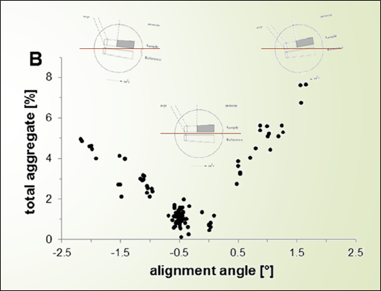
AUC is an orthogonal technique to SEC widely employed for aggregate characterization as shown in Figure 3. It is often used to study protein higher-order structure and assembly mechanism. Despite several advantages, SEC is not feasible when a direct analysis of the sample is required in its native buffer since SEC is conducted in a mobile phase which is significantly different from the sample matrix. In addition to this, the initial sample concentration is greatly reduced during injection. In such cases, AUC is the preferable method for aggregate analysis or it can be used as a confirmatory technique along with SEC (Chaturvedi et al., 2020). AUC experiments can be done in two ways: Sedimentation equilibrium (SE-AUC) quantifies the equilibrium concentration distribution of macromolecules, while Sedimentation velocity (SV-AUC) monitors real-time sedimentation of macromolecules. SV-AUC is the preferred method to study aggregation since it works well in different types of solutions including in the matrix of drug substance and drug product without any matrix interaction. However, there are a few limitations to this technique such as low throughput, the need for experienced analysts, potential gradient formation with certain excipients, and a restricted dynamic detection range (Bou-Assaf et al., 2022). The most commonly used method to assess protein aggregation by SV-AUC is sedimentation coefficient distribution analysis using SEDFIT software. This method estimates the range of sedimentation coefficients in a mixture of proteins, buffers, and excipients alongside providing an insight into the mass of aggregates in a protein product (Healey et al., 2017).
|
|
|
Figure 3. Alignment of the centrifuge cells in the rotor is critical to accurate aggregate quantitation (Bou-Assaf et al., 2022). |
Conclusion
In summary, detailed monoclonal antibody characterization, including identity, heterogeneity, impurities, and stability, is essential for the drug development and manufacturing process. A broad range of analytical techniques are employed to characterize mAbs at different stages of its development and manufacturing. Adherence to ICH and other applicable regulatory guidelines is crucial for releasing biopharmaceutical drugs in the market.
This review has specifically focused on the critical aspect of mAb aggregation, which leads to the transformation of native proteins into higher molecular weight species. Aggregate detection is crucial as it results in increased immunogenicity, potentially leading to adverse reactions in patients. Various analytical techniques such as SEC, AUC, UV-visible, Fluorescence spectroscopy, and DLS have been studied extensively to detect aggregation. Each technique has its advantages and limitations; therefore, a combination of techniques is recommended for a more comprehensive understanding. The insights gained from these techniques contribute to the development of safe and high-quality biopharmaceutical drugs. As the next-generation technologies emerge, mAb characterization will continue to advance, becoming a vast area to explore within the realms of biopharmaceutical and biologics division.
Acknowledgments: None
Conflict of interest: None
Financial support: None
Ethics statement: None
Bansal, R., Gupta, S., & Rathore, A. S. (2019). Analytical platform for monitoring aggregation of monoclonal antibody therapeutics. Pharmaceutical Research, 36(11), 1-11. doi:10.1007/s11095-019-2690-8
Bou-Assaf, G. M., Budyak, I. L., Brenowitz, M., Day, E. S., Hayes, D., Hill, J., Majumdar, R., Ringhieri, P., Schuck, P., & Lin, J. C. (2022). Best practices for aggregate quantitation of antibody therapeutics by sedimentation velocity analytical ultracentrifugation. Journal of Pharmaceutical Sciences, 111(7), 2121-2133. doi:10.1016/j.xphs.2021.12.023
Chaturvedi, S. K., Parupudi, A., Juul-Madsen, K., Nguyen, A., Vorup-Jensen, T., Dragulin-Otto, S., Zhao, H., Esfandiary, R., & Schuck, P. (2020). Measuring aggregates, self-association, and weak interactions in concentrated therapeutic antibody solutions. mAbs, 12(1), 1810488. doi:10.1080/19420862.2020.1810488
Harding S. E. (2022). Biophysical Reviews' "meet the editors series"-a profile of Steve Harding's career in macromolecular hydrodynamics. Biophysical Reviews, 14(3), 605-610. doi:10.1007/s12551-022-00963-5
Healey, J. F., Parker, E. T., & Lollar, P. (2018). Identification of aggregates in therapeutic formulations of recombinant full-length factor VIII products by sedimentation velocity analytical ultracentrifugation. Journal of Thrombosis and Haemostasis: JTH, 16(2), 303–315. doi:10.1111/jth.13917
Ilochonwu, B. C., Urtti, A., Hennink, W. E., & Vermonden, T. (2020). Intravitreal hydrogels for sustained release of therapeutic proteins. Journal of controlled Release: Official Journal of the Controlled Release Society, 326, 419-441. doi:10.1016/j.jconrel.2020.07.031
Jun, B. M., Kim, S., Rho, H., Park, C. M., & Yoon, Y. (2020). Ultrasound-assisted Ti3C2Tx MXene adsorption of dyes: Removal performance and mechanism analyses via dynamic light scattering. Chemosphere, 254, 126827. doi:10.1016/j.chemosphere.2020.126827
Kaur, H. (2021). Physicochemical characterization of monoclonal antibodies. In Monoclonal Antibodies (pp. 31-63). Academic Press.
Laptoš, T., & Omersel, J. (2018). The importance of handling high-value biologicals: Physico-chemical instability and immunogenicity of monoclonal antibodies. Experimental and Therapeutic Medicine, 15(4), 3161-3168. doi:10.3892/etm.2018.5821
Le Basle, Y., Chennell, P., Tokhadze, N., Astier, A., & Sautou, V. (2020). Physicochemical stability of monoclonal antibodies: A review. Journal of Pharmaceutical Sciences, 109(1), 169-190. doi:10.1016/j.xphs.2019.08.009
Li, W., Prabakaran, P., Chen, W., Zhu, Z., Feng, Y., & Dimitrov, D. S. (2016). Antibody aggregation: Insights from sequence and structure. Antibodies (Basel, Switzerland), 5(3), 19. doi:10.3390/antib5030019
Loa, J. D. A., Cruz-Rodríguez, I. A., & Rojas-Avelizapa, N. G. (2023). Colorimetric detection of metals using CdS-NPs synthesized by an organic extract of aspergillus Niger. Applied Biochemistry and Biotechnology, 195(7), 4148-4163. doi:10.1007/s12010-023-04341-z
Maiti, R., Patel, B., Patel, N., Patel, M., Patel, A., & Dhanesha, N. (2023). Antibody drug conjugates as targeted cancer therapy: past development, present challenges and future opportunities. Archives of Pharmacal Research, 46(5), 361-388. doi:10.1007/s12272-023-01447-0
Nobbmann, U., Connah, M., Fish, B., Varley, P., Gee, C., Mulot, S., Chen, J., Zhou, L., Lu, Y., Shen, F., et al. (2007). Dynamic light scattering as a relative tool for assessing the molecular integrity and stability of monoclonal antibodies. Biotechnology & Genetic Engineering Reviews, 24(1), 117-128. doi:10.1080/02648725.2007.10648095
Paul, A. J., Bickel, F., Röhm, M., Hospach, L., Halder, B., Rettich, N., Handrick, R., Herold, E. M., Kiefer, H., & Hesse, F. (2017). High-throughput analysis of sub-visible mAb aggregate particles using automated fluorescence microscopy imaging. Analytical and Bioanalytical Chemistry, 409(17), 4149-4156. doi:10.1007/s00216-017-0362-2
Ranadive, P., Bedi, S., Bhalla, A. S., & Shameem, M. (2023). Water loss from silicone tubing and effect on protein concentration during drug product manufacturing. European Journal of Pharmaceutics and Biopharmaceutics: Official Journal of Arbeitsgemeinschaft fur Pharmazeutische Verfahrenstechnik e.V, 185, 116-125. doi:10.1016/j.ejpb.2022.12.015
Sadighi, R., de Kleijne, V., Wouters, S., Lubbers, K., Somsen, G. W., Gargano, A. F. G., & Haselberg, R. (2024). Online multimethod platform for comprehensive characterization of monoclonal antibodies in cell culture fluid from a single sample injection - Intact protein workflow. Analytica Chimica Acta, 1287, 342074. doi:10.1016/j.aca.2023.342074
Saldanha, M., Shelar, A., Patil, V., Warke, V. G., Dandekar, P., & Jain, R. (2023). A case study: Correlation of the nutrient composition in Chinese Hamster Ovary cultures with cell growth, antibody titre and quality attributes using multivariate analyses for guiding medium and feed optimization in early upstream process development. Cytotechnology, 75(1), 77-91. doi:10.1007/s10616-022-00561-z
São Pedro, M. N., Klijn, M. E., Eppink, M. H., & Ottens, M. (2022). Process analytical technique (PAT) miniaturization for monoclonal antibody aggregate detection in continuous downstream processing. Journal of Chemical Technology & Biotechnology, 97(9), 2347-2364. doi:10.1002/jctb.6920
Sats, A., Kaart, T., & Jõudu, I. (2024). Bovine colostrum casein: Post-partum dynamics of micelle size, content, and associated traits. International Dairy Journal, 148(3), 105791. doi:10.1016/j.idairyj.2023.105791
Sats, A., Kaart, T., Poikalainen, V., Aare, A., Lepasalu, L., Andreson, H., & Jõudu, I. (2020). Bovine colostrum whey: Postpartum changes of particle size distribution and immunoglobulin G concentration at different filtration pore sizes. Journal of Dairy Science, 103(8), 6810-6819. doi:10.3168/jds.2019-17604
Sharma, A., Beirne, J., Khamar, D., Maguire, C., Hayden, A., & Hughes, H. (2023). Evaluation and screening of biopharmaceuticals using multi-angle dynamic light scattering. AAPS PharmSciTech, 24(4), 84. doi:10.1208/s12249-023-02529-4
Van der Kant, R., Karow-Zwick, A. R., Van Durme, J., Blech, M., Gallardo, R., Seeliger, D., Aßfalg, K., Baatsen, P., Compernolle, G., Gils, A., et al. (2017). Prediction and reduction of the aggregation of monoclonal antibodies. Journal of Molecular Biology, 429(8), 1244-1261. doi:10.1016/j.jmb.2017.03.014
Wang, F. A. S., Fan, Y., Chung, W. K., Dutta, A., Fiedler, E., Haupts, U., Peyser, J., & Kuriyel, R. (2024). Evaluation of mild pH elution protein A resins for antibodies and Fc-fusion proteins. Journal of Chromatography A, 1713(1), 464523. doi:10.1016/j.chroma.2023.464523
Xu, M. N., Zhong, M. Z., Feng, S. N., Xu, Y. Q., Peng, X. M., Zeng, K., & Huang, X. W. (2024). Production of recombinant HPV11/16 E6/E7-MBP-His6 fusion proteins and their potential to induce cytokine secretion by immune cells in peripheral blood. Virology Journal, 21(1), 10. doi:10.1186/s12985-023-02281-y