Collagen Hydrolysates in the Prevention and Treatment of Arthritis
Anna Vladimirovna Kryuchkova, Runa Usmanovna Tunguzbieva, Kumira Sultanovna Tokaeva, Adam Aiupovich Isaev, Layla Ruslanovna Elmaeva*, Viktor Vasilievich Mikhailenko
Abstract
The present review examines the role of collagen hydrolysates in the treatment and prevention of joint diseases. Collagen is the main structural protein of bones, cartilage, ligaments, and tendons in joints. Hydrolyzed collagen, as a source of specific amino acids, can be a building material for the formation of collagen fibrils in connective tissues. Enzymatic hydrolysis of chicken cartilage made it possible to obtain a complex of collagen type II hydrolysate and glycosaminoglycans. In clinical studies, hydrolysates of cutaneous collagen, type II cartilaginous collagen, and type II non-denatured cartilaginous collagen were tested. The use of collagen hydrolysates for a long time (5-6 months) allowed for improving the functioning of the knee joint in patients not only with mild but also with severe osteoarthritis. Nutraceuticals from type II collagen hydrolysates in combination with vitamin C more effectively affect the formation of collagen fibrils, and proteoglycans in the cartilage matrix and thus can affect the increase in joint mobility. Nutriciology, as a branch of modern biopharmaceutics, represents its new direction and explores possible mechanisms and ways to prevent human diseases.
Keywords: Arthritis, Nutraceuticals, Medicine, Joint, Collagen
Introduction
Cartilage diseases: arthritis and arthrosis of joints and destruction of intervertebral discs in the spine are common chronic diseases (Gabay & Clouse, 2016). By pharmacological agents, (steroids and other drugs) diseases of the musculoskeletal system are difficult to treat (Hollister, 1992). The medications used are mainly aimed at reducing pain and increasing joint mobility. Therefore, medical institutions and patients are looking for non-standard methods of eliminating health disorders (Bledzhyants et al., 2019; Mezhidov et al., 2021). In recent years, along with traditional pharmacotherapy, polypharmaceuticals, and biopharmaceuticals have been developing (Pfeifer et al., 2021). Clinical nutritionology is one of the new directions of biopharmaceutics (Lobo & Deutz, 2020). By definition, accepted in the USA and the UK, a nutraceutical is a food with pharmaceutical properties. One of the problems of the rational search for natural medicines capable of providing biopharmaceutical and polypharmaceutical effects can be solved by the development of nutraceuticals from connective tissue components (Deal & Moskowitz, 1999). Nutraceuticals containing collagen hydrolysate, glucosamine, chondroitin sulfate, hyaluronic acid, vitamin C, and other substances have become widespread (Bazhenova et al., 2021). Glycosaminoglycans – chondroprotectors – have long been used in diseases of the musculoskeletal system (Danilov & Grigorenko, 2015). In this review, we consider collagen as the main protein of connective tissues, the fibrils of which strengthen the matrix of tissues in the joints. By applying collagen hydrolysates, patients can prevent or alleviate the symptoms of diseases. The purpose of this review is to analyze nutraceuticals derived from collagen hydrolysates, as well as to evaluate the effectiveness of their action in the prevention and treatment of joint diseases.
Structure of Connective Tissues of Joints
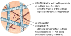
Such a complex organ as a joint consists of bones, cartilage, ligaments, tendons, and vitreous (Figure 1) (Ralphs & Benjamin, 1994). Cartilage tissue is localized in those areas of the musculoskeletal system that experience the greatest mechanical stress. Changes in joints in diseases begin primarily in hyaline cartilage and then spread to the bone and other tissues (Jeffrey & Watt, 2003).
The connective tissues of the joints are formed from specialized cells and extracellular matrix. The matrix contains collagen and elastin fibrils, proteoglycans, and glycoproteins. The mechanical strength of the matrix is determined by the rigid structure of collagen and elastin fibrils (Theocharis et al., 2016). Collagen is the main structural protein of connective tissues. Figure 1 shows the main components of cartilage tissue and describes their role.
Fibrils are formed from collagens of types I, II, and III and their complexes with minor collagens of types IX, X, XI, XII, and XIV. Type II collagen predominates in cartilage (Luo et al., 2017). Type VI collagen fibrils connect the main fibrils. Collagen fibrils in the matrix form a three-dimensional network. Proteoglycans and flexible fibrils of small diameter, but containing a large number of covalent cross-links, give elasticity to tissues (Silver et al., 2021). Proteoglycans and glycoproteins fill the space between fibrils and cells. Glycoproteins, binding to receptors on the surface of chondrocytes, interact with the matrix with cells. Proteoglycans are formed from complexes of aggrecans, binding protein, and hyaluronic acid. The composition of aggrecans includes glycosaminoglycans: chondroitin-4-sulfate, chondroitin-6-sulfate, and keratan-sulfate (Ren et al., 2021). The specific structure of the matrix provides both biomechanical properties of cartilage and promotes its nutrition by diffusion. It is known that there is no circulatory system in hyaline cartilage (Jeffrey & Watt, 2003). Nevertheless, the interstitial water, its large amount, and the substances dissolved in it provide an intensive metabolism. Proteoglycans, having cationic and anionic properties, regulate the physicochemical properties of the matrix. Changes in the content of collagen proteins, proteoglycans, glycosaminoglycans, metal ions, and water disrupt the structure of cartilage tissue, leading to diseases. Pathological processes in arthrosis are accompanied by reduced concentrations of collagen and glycosaminoglycans (Eyre, 2002). The structure of collagen fibrils in the joints is weakened in the hereditary disease hypermobility of the joints, in which the skin also becomes thinner (Wolf et al., 2011). Violations of the matrix structure and the size of collagen fibrils in such different tissues as cartilage and skin occur in the absence of type VI collagen, a deficiency of salts and vitamins (Kuo et al., 1995). The replacement of the missing components of the matrix in the human body can be carried out using therapeutic agents of preventive action, as well as nutraceuticals. Chondroprotective drugs are hyaluronic acid and sulfated glycosaminoglycans (Sukhikh et al., 2020). They are isolated from proteoglycan complexes contained in the tissues of vertebrates and hydrobionts. The macromolecules of connective tissues embedded by nature in the human and animal bodies can be drugs of polypharmaceutical and biopharmaceutical action.
|
|
|
Figure 1. The role of components in cartilage tissue |
Production of Collagen Hydrolysates and Nutraceuticals Based on Them
Even though collagen is the main protein of joints, collagen nutrients have been used relatively recently for the treatment of arthritis and arthrosis. Collagen enters the human body with food after its heat treatment. the denatured type of collagen is medicinal and food gelatin (Liu et al., 2015). Collagen proteins and gelatin under the action of enzymes of the gastrointestinal tract can be broken down into amino acids and polypeptides. The large length of collagen molecules and fibrils, which have a relatively high molecular weight, prevents their effective digestion (Olsen et al., 2003). Collagen hydrolysates containing a set of amino acids and polypeptides are more readily available for assimilation in the human body. Amino acids entering the blood accumulate in connective tissues and cells. Hydrolyzed collagen, as a source of specific amino acids, can be a building material for the biosynthesis of the matrix of these tissues (León-López et al., 2019).
Pharmaceutical gelatin was obtained from extracts of skin tissue. To increase the absorption of gelatin, its enzymatic hydrolysis was additionally carried out (Borges et al., 2016).
An urgent task is to obtain collagen hydrolysates with a high content of free amino acids and low molecular weight polypeptides. Enzymatic hydrolysis of collagen is a necessary stage in the creation of effective nutraceuticals and the production of nutrients with regulated characteristics (Skov et al., 2019).
The degree of hydrolysis (DH) is the most significant characteristic of the depth of hydrolysis. The DH index correlates with the distribution of molecular chain lengths and with the molecular weight of peptides. The molecular parameters of hydrolysates vary depending on the type of animal and the conditions of tissue hydrolysis. Thinner fish skin (cod and other species) promotes the formation of collagen hydrolysates of a higher degree of hydrolysis when compared with hydrolysates of hydrobionts (trepangs) (Blanco et al., 2017). Tissue hydrolysis was studied under the influence of different enzymes, and the duration of the enzyme, as well as depending on temperature, pH, and enzyme/substrate ratio. Optimization of the conditions of enzymatic hydrolysis allows for an increase in the degree of collagen hydrolysis.
Hydrolyzate of collagen from the skin of cattle "Collamine-80" was obtained under the influence of enzymes in the pancreas of pigs. "Collamine-80" contains amino acids, dipeptides, tripeptides, and polypeptides. Based on "Collamine-80" in Russia, the nutraceuticals "Collagen ULTRA" and "Collagen-S" were developed. Their main component is the hydrolyzate of collagen types I and III. However, the skin tissue hydrolyzate used in all nutraceuticals does not contain a complete set of amino acids specific for cartilage fibrils and joints. Based on the difference in the amino acid composition of type I, II, and III collagens, we suggest that nutrients from skin collagens may have a weaker effect on the formation of strong fibrils in the tissues of the musculoskeletal system (Mafi et al., 2012).
From chicken cartilage, using the proteolytic enzymes papain, ficin, and bromelain, type II collagen hydrolysates were obtained in the USA. The molecular weight of the peptides varies from 50 to 10,000 d. Along with type II collagen, BioCell Collagen II hydrolyzate contains glycosaminoglycans: hyaluronic acid, and chondroitin sulfate. Based on these hydrolysates, nutraceuticals were developed with a set of other components: JointFlex Complete, Flex-a-Min, and Flex-A-Min Triple Strength.
Experiments in Cell Culture
It was shown that in chondrocytes in vitro after 11 days of cultivation in a medium with type II collagen hydrolyzate, the number of amino acids increased by 2.5 times compared to the control (medium without collagen hydrolyzate). Stimulation of collagen formation from amino acids in cells is regulated by enzymes whose activity is influenced by vitamins and metal cations (Abujamel, 2022). Synthesis of collagen and its fibril formation is a complex multi-stage process. Biosynthesis takes place in specialized cells (chondrocytes in cartilage), and fibril formation occurs near the cell surface (Bahshwan, 2022). Hydroxylation reactions make it possible to modify collagen molecules. Enzymes catalyze hydroxylation: prolyl-4-hydroxylase and lysyl-5-hydroxylase to convert proline to hydroxyproline, and lysine to oxylysine. Enzymes are active if iron is in the divalent form, which is provided by ascorbic acid (vitamin C). A feature of high-strength collagen fibrils is the high content of amino acid residues with aldehyde groups. Normally, lysine and oxylysine form their aldehyde forms, allisin, and oxyallysin, which are involved in the formation of strong covalent bonds between collagen molecules. Therefore, nutraceuticals from collagen hydrolysates in combination with ascorbic acid (vitamin C) have a more effective effect on fibril formation.
The cartilage of the joints accumulates ascorbic acid. In chondrocytes, ascorbic acid and dihydroascorbate are translocated via sodium-dependent vitamin C transport (SVCT-2) and glucose transport GLUT 1, respectively. In vitro, ascorbate and ascorbic acid stimulated the synthesis of type I and II collagen, proteoglycans, and aggrecans in joint chondrocytes (Schendrigin et al., 2022). An in vivo study in STR/ort mice (which developed osteoarthritis) showed that long-term administration of collagen hydrolyzate can reduce the degenerativeness of cartilage tissue affected by osteoarthritis and delay its development (Rzhepakovsky et al., 2021). Table 1 shows inducers and inhibitors of collagen synthesis.
Table 1. Inducers and inhibitors of collagen synthesis
|
Synthesis inhibitors |
Synthesis inductors |
|
Epidermal growth factor |
Insulin-like growth factor I |
|
Fibroblast growth factor |
Transformative growth factor β1 |
|
Main inflammatory mediators (IL-1, TNFα and interferon-γ) |
|
|
Fibroblast cell surface integrins – receptor α1β1 |
Fibroplast cell surface integrins – receptor α1β1 |
|
Platelet growth factor |
Platelet growth factor |
|
Hormones - gluccorticosteroids |
Hormones - insulin, progesterone, androgens |
|
Deficiency of enzymes, cofactors |
|
Clinical Studies
After clinical trials of chondroprotectors, both positive and negative results from the use of chondroitin sulfate and hyaluronic acid were observed. The insignificant effect of these chondroprotectors on pain reduction in patients has also been established. The inconsistency of the observed difference is apparently due to the different sources and methods of obtaining chondroprotectors (Shavlovskaya et al., 2020). If along with glycosaminoglycans, collagen proteins are included in the composition of the drug, the drug may have a stimulating effect. The lack of collagen proteins is compensated by taking nutraceuticals (Asiwe et al., 2022; Chakravert & Bondyopadhyay, 2022; Seal et al., 2022; Subbaiah et al., 2022)
The effect of pharmaceutical collagen hydrolysate on metabolism in patients with osteoarthritis was tested (Moskowitz, 2000). Patients with osteoarthritis of the knee joint took 10 g of pharmaceutical collagen hydrolysate or 12 g of lactose (placebo) daily for 24 weeks. Clinical studies in medical centers in three countries have revealed an improvement in joint function and the advantage of treatment with pharmaceutical collagen hydrolysate over placebo only in Germany. No statistically reliable results were obtained in either the US or the UK.
The specificity of connective tissue for obtaining collagen hydrolysates and more effective treatment of joints has been determined. After the use of type II collagen hydrolysate for 24 weeks in a group of athletes – healthy people, but with intraarticular pain - an increase in joint mobility, a decrease in pain syndrome, and a decrease in dependence on analgesics were observed. In patients with primary osteoarthritis, under the same conditions of receiving collagen hydrolysate, the knee joint function improved according to the visual analog scale and WOMAC. In patients with symptoms of mild osteoarthritis of the knee joint, who used collagen hydrolysate (10 g/day) with calcium (300 mg/day) and vitamin C (60 mg/day) for 14 weeks, the functional mobility of the knee increased according to isometric and isokinetic tests. Also, higher results were observed in patients with severe forms of osteoarthritis.
Non-denatured type II collagen was used to treat rheumatoid arthritis. The study was carried out at Harvard University in 1993. The results were obtained that non-denatured type II collagen can affect joint mobility and pain reduction in them, as well as improve the functional state of patients with osteoarthritis (Moskowitz, 2000). The potential mechanism of action of non-denatured type II collagen is probably due to the "retraining" of T cells for immune resistance. Intact type II collagen entering the gastrointestinal tract re-creates antigenic interactions with tree cells and regulatory T cells in the lymph of intestinal tissue. Regulatory T cells secrete cytokines, such as interleukin-10 and transforming growth factor, which inhibits the immune response to antigens (type II collagen). Cytokines can affect the reduction of the immune response to type II collagen inside the articular cartilage and thus prevent an anti-inflammatory acute reaction to articular cartilage in conditions of arthritis. Considering the complementary mechanism of action of non-denatured type II collagen in addition to its effectiveness, it can be considered a complex-acting supplement and can be taken at 40 mg daily (Anastasova et al., 2022; Jallepalli et al., 2022; Mane & Kanase, 2022; Zibi et al., 2022).
Nutritionology in the Prevention of Joint Diseases
Currently, the objectives of treating patients with arthritis and arthrosis are to increase joint mobility and reduce pain. Joint mobility is determined by the optimal size of collagen fibrils and proteoglycans. It is known that in cartilage matrix renewal occurs after a year (Strecanska et al., 2022). Therefore, the long-term and systematic use of a complex of specific amino acids and glycosaminoglycans in type II collagen hydrolysates makes it possible to restore and strengthen the structure of human joint tissues. The recommended dose is 10 g of collagen hydrolysate per day. Nutraceuticals based on collagen hydrolysate effectively affect both the early stage of joint diseases and the prevention of such diseases. Chronic human diseases due to connective tissue dysplasia are laid at the embryonic stage of development and /or at the growth stages of children and adolescents. Nutraceutical supplements made of collagen amino acids help weakened children strengthen cartilage, bone, and other connective tissues (Khatri et al., 2021). The use of nutraceuticals from collagen hydrolysates in combination with glycosaminoglycans, vitamins, and metal cations can stimulate the biosynthesis of macromolecules in cells and the structure of the extracellular matrix, disrupted as a result of diseases (Cardoso et al., 2014; Mobasheri et al., 2021; Blinov et al., 2022). If the action of modern medicines is aimed at reducing inflammatory and painful symptoms during the disease or its exacerbation, then nutraceuticals have an advantage in preventing diseases (Alanazi et al., 2019; Mostafavi et al., 2019; Son et al., 2021).
Conclusion
Taking into account that collagen is the main structural protein of bones, cartilage, ligaments, and tendons in joints, the role of collagen hydrolysates in the treatment and prevention of joint diseases is considered. to obtain hydrolysates with a high content of amino acids, a biocatalytic approach and optimization of enzymatic hydrolysis are effective. The data presented in the review show that collagen hydrolysates can alleviate the symptoms of joint diseases. However, the role of nutrition in reducing the development of the disease remains poorly understood. Research on the treatment and prevention of connective tissue diseases using nutraceuticals is at an early stage of development. Only a few types of collagen hydrolysate have been obtained and tested. Moreover, the specificity of type II collagen hydrolysate from cartilage tissue has been determined to improve the functioning of joints. Since the increase in joint mobility can be regulated by the size of collagen fibrils and proteoglycans, it will be necessary to determine the relationship between the composition of collagen hydrolysates and the structure of the matrix. If the pharmacological method is based on the study of one molecule / one target, then nutritionology is a more holistic type of methodology: many ingredients / multiple targets. Nutriciology, as a branch of modern biopharmaceutics, represents its new direction and explores possible mechanisms and ways to prevent human diseases.
Acknowledgments: The authors are thankful to colleagues from Stavropol State Agrarian University for consultancy dring literature review.
Conflict of interest: None
Financial support: None
Ethics statement: None
Abujamel, T. S. (2022). Understanding the mechanisms of bacterial antimicrobial resistance within biofilms. International Journal of Pharmaceutical and Phytopharmacological Research, 12(1), 17-24. doi:10.51847/o5Bt4kEqyT
Abujamel, T. S. (2022). Understanding the mechanisms of bacterial antimicrobial resistance within biofilms. International Journal of Pharmaceutical and Phytopharmacological Research, 12(1), 17-24. doi:10.51847/o5Bt4kEqyT
Alanazi, A. M., Alotaibi, H. D., Alahmari, S. A., Almutairi, A. K., Babakr, S. A. A., Abdrabalnabi, H. A. A., Ali, A. A., Shamat, R. A. A. A., Husain, Z. A., Abdulla, A. H. A., et al. (2019). Hip bone fracture diagnosis and management. Archives of Pharmacy Practice, 10(4), 29-32.
Anastasova, L., Ivanovska, T. P., Ancevska, A., Petkovska, R., & Petrushevska-Tozi, L. (2022). Applıcatıon of experımental desıgn approach in optımızatıon of qualıty parameters of calcıum- and magnesıum-enrıched mılk. International Journal of Pharmaceutical and Phytopharmacological Research, 12(1), 7-16. doi:10.51847/MtCiwMuW5D
Asiwe, N., Asiwe, J. N., Asiwe, T. N., & Asiwe, P. C. (2022). Awareness of COVID-19 and its vaccine acceptability among young adult population of Agbor, Delta State, Nigeria. International Journal of Pharmaceutical and Phytopharmacological Research, 12(2), 24-29. doi:10.51847/tvmGc5ytyZ
Bahshwan, S. M. (2022). Effect of different levels of (V. Agnus-Castus) on the immunological & histopathological changes in hepatic rats. International Journal of Pharmaceutical and Phytopharmacological Research, 12(1), 38-43. doi:10.51847/eWbaaSHAXa
Bazhenova, A. A., Guryanova, N. I., Guryanov, G. S., Alieva, H. A. V., Kachmazova, D. T., Khripunova, A. A., & Povetkin, S. N. (2021). In-Vitro study of the properties of components for the synthesis of sorbent for low-density lipoprotein apheresis. Pharmacophore, 12(3), 37-41. doi:10.51847/BsjhKFW0Kd
Blanco, M., Vázquez, J. A., Pérez-Martín, R. I., & Sotelo, C. G. (2017). Hydrolysates of fish skin collagen: An opportunity for valorizing fish industry byproducts. Marine Drugs, 15(5), 131. doi:10.3390/md15050131
Bledzhyants, G. A., Mishvelov, A. E., Nuzhnaya, K. V., Anfinogenova, O. I., Isakova, J. A., Melkonyan, R. S., Hite, G. Y., Suprunchuk, V. E., Makova, A. V., Popov, A. N., et al. (2019). The effectiveness of the medical decision-making support system" electronic clinical pharmacologist" in the management of patients therapeutic profile. Pharmacophore, 10(2), 76-81.
Blinov, A. V., Maglakelidze, D. G., Yasnaya, M. A., Gvozdenko, A. A., Blinova, A. A., Golik, A. B., Slyadneva, K. S., & Pirogov, M. A. (2022). Synthesis of selenium nanoparticles stabilized by quaternary ammonium compounds. Russian Journal of General Chemistry, 92(3), 424-429. doi:10.1134/S1070363222030094
Borges, J. G., Silva, A. G., Cervi-Bitencourt, C. M., Vanin, F. M., & Carvalho, R. A. D. (2016). Lecithin, gelatin and hydrolyzed collagen orally disintegrating films: Functional properties. International Journal of Biological Macromolecules, 86, 907-916. doi:10.1016/j.ijbiomac.2016.01.089
Cardoso, V. S., Quelemes, P. V., Amorin, A., Primo, F. L., Gobo, G. G., Tedesco, A. C., Mafud, A. C., Mascarenhas, Y. P., Corrêa, J. R., Kuckelhaus, S. A., et al. (2014). Collagen-based silver nanoparticles for biological applications: Synthesis and characterization. Journal of Nanobiotechnology, 12, 1-9. doi:10.1186/s12951-014-0036-6
Chakraverty, R., & Bondyopadhyay, J. (2022). Recent insights into the association between stress, anxiety and hypertension in adults: a systematic review. International Journal of Pharmaceutical and Phytopharmacological Research, 12(2), 12-17. doi:10.51847/Bug18QDArj
Danilov, A. B., & Grigorenko, N. V. (2015). An antinociceptive effect of chondroprotectors: A myth or a reality? Zhurnal nevrologii i psikhiatrii imeni SS Korsakova, 115(9), 84-89. doi:10.17116/jnevro20151159184-89
Deal, C. L., & Moskowitz, R. W. (1999). Nutraceuticals as therapeutic agents in osteoarthritis: The role of glucosamine, chondroitin sulfate, and collagen hydrolysate. Rheumatic Disease Clinics of North America, 25(2), 379-395. doi:10.1016/s0889-857x(05)70074-0
Eyre, D. (2002). Collagen of articular cartilage. Arthritis Research, 4(1), 30-35. doi:10.1186/ar380
Gabay, O., & Clouse, K. A. (2016). Epigenetics of cartilage diseases. Joint Bone Spine, 83(5), 491-494. doi:10.1016/j.jbspin.2015.10.004
Hollister, J. R. (1992). The untoward effects of steroid treatment on the musculoskeletal system and what to do about them. Journal of Asthma, 29(6), 363-368. doi:10.3109/02770909209044799
Jallepalli, V. R., Thalla, S., Gavini, S. B., Tella, J. D., Kanneganti, S., Yemineni, G., & Nadendla, R. R. (2022). Impact of patient education on quality of life in gastroesophageal reflux disease. International Journal of Pharmaceutical and Phytopharmacological Research, 12(1), 25-28. doi:10.51847/dAJecTWofD
Jeffrey, D. R., & Watt, I. (2003). Imaging hyaline cartilage. The British Journal of Radiology, 76(911), 777-787. doi:10.1259/bjr/51504520
Khatri, M., Naughton, R. J., Clifford, T., Harper, L. D., & Corr, L. (2021). The effects of collagen peptide supplementation on body composition, collagen synthesis, and recovery from joint injury and exercise: A systematic review. Amino Acids, 53(10), 1493-1506. doi:10.1007/s00726-021-03072-x
Kuo, H. J., Keene, D. R., & Glanville, R. W. (1995). The macromolecular structure of type‐VI collagen: Formation and stability of filaments. European Journal of Biochemistry, 232(2), 364-372.
León-López, A., Morales-Peñaloza, A., Martínez-Juárez, V. M., Vargas-Torres, A., Zeugolis, D. I., & Aguirre-Álvarez, G. (2019). Hydrolyzed collagen—Sources and applications. Molecules, 24(22), 4031. doi:10.3390/molecules24224031
Liu, D., Nikoo, M., Boran, G., Zhou, P., & Regenstein, J. M. (2015). Collagen and gelatin. Annual Review of Food Science and Technology, 6, 527-557. doi:10.1146/annurev-food-031414-111800
Lobo, D. N., & Deutz, N. E. (2020). Publication governance in clinical nutrition. Clinical Nutrition, 39(1), 1-4. doi:10.1016/j.clnu.2019.11.010
Luo, Y., Sinkeviciute, D., He, Y., Karsdal, M., Henrotin, Y., Mobasheri, A., Önnerfjord, P., & Bay-Jensen, A. (2017). The minor collagens in articular cartilage. Protein & Cell, 8(8), 560-572. doi:10.1007/s13238-017-0377-7
Mafi, P., Hindocha, S., Mafi, R., & Khan, W. (2012). Evaluation of biological protein-based collagen scaffolds in cartilage and musculoskeletal tissue engineering-A systematic review of the literature. Current Stem Cell Research & Therapy, 7(4), 302-309. doi:10.2174/157488812800793045
Mane, S. T., & Kanase, D. G. (2022). Recent developments in chiral stationary phases: a mini-review. International Journal of Pharmaceutical and Phytopharmacological Research, 12(1), 44-53. doi:10.51847/ywEtn2HoHl
Mezhidov, B. S., Belyaeva, A. A., Kh, S. M., Bimarzaev, S., Bektashev, A., Shekhshebekova, A. M., & Dzgoeva, M. G. (2021). Prospects for creating 3D models of internal organs based on computer and magnetic resonance imaging images in emergency surgery and resuscitation. Pharmacophore, 11(1), 8-14. doi:10.51847/3TLcii4n42
Mobasheri, A., Mahmoudian, A., Kalvaityte, U., Uzieliene, I., Larder, C. E., Iskandar, M. M., Kubow, S., Hamdan, P. C., de Almeida, C. S., Favazzo, L. J., et al. (2021). A white paper on collagen hydrolyzates and ultrahydrolyzates: Potential supplements to support joint health in osteoarthritis? Current Rheumatology Reports, 23, 1-15. doi:10.1007/s11926-021-01042-6
Moskowitz, R. W. (2000). Role of collagen hydrolysate in bone and joint disease. In Seminars in arthritis and rheumatism (Vol. 30, No. 2, pp. 87-99). WB Saunders. doi:10.1053/sarh.2000.9622
Mostafavi, Z., Houshyar, J., Aliasgarzadeh, A., Aghamohammadzadeh, N., Sadra, V., Khani, M., Najafipour, M., & Najafipour, F. (2019). Comparison of the bone mineral density between acromegaly patients and healthy individuals. Archives of Pharmacy Practice, 10(4), 15-21.
Olsen, D., Yang, C., Bodo, M., Chang, R., Leigh, S., Baez, J., Carmichael, D., Perälä, M., Hämäläinen, E. R., Jarvinen, M., et al. (2003). Recombinant collagen and gelatin for drug delivery. Advanced Drug Delivery Reviews, 55(12), 1547-1567. doi:10.1016/j.addr.2003.08.008
Pfeifer, B. A., Zhang, G., & Li, D. (2021). Editorial overview: Pharmaceutical biotechnology. Current Opinion in Biotechnology, 69, vi-viii. doi:10.1016/j.copbio.2021.06.001
Ralphs, J. R., & Benjamin, M. (1994). The joint capsule: Structure, composition, ageing and disease. Journal of Anatomy, 184(Pt 3), 503-509.
Ren, Q., Wang, J., Liu, C., Meng, L. X., Qian, R. K., Gao, H. J., Qin, W., Zhou, C. J., Qiao, S., Wang, H. Y., et al. (2021). Exploring the sulfate patterns of chondroitin sulfate/dermatan sulfate and keratan sulfate in human pancreatic cancer. Journal of Pharmaceutical and Biomedical Analysis, 205, 114339. doi:10.1016/j.jpba.2021.114339
Rzhepakovsky, I., Anusha Siddiqui, S., Avanesyan, S., Benlidayi, M., Dhingra, K., Dolgalev, A., Enukashvily, N., Fritsch, T., Heinz, V., Kochergin, S., et al. (2021). Anti‐arthritic effect of chicken embryo tissue hydrolyzate against adjuvant arthritis in rats (X‐ray microtomographic and histopathological analysis). Food Science & Nutrition, 9(10), 5648-5669. doi:10.1002/fsn3.2529
Schendrigin, I. N., Timchenko, L. D., Rzhepakovsky, I. V., Avanesyan, S. S., Sizonenko, M. N., Grimm, W. D., Povetkin, S. N., & Piskov, S. I. (2022). Clinical and pathogenetic significance of amylase level and microtomographic index of synovial fluid in various joint lesions. Современные технологии в медицине, 14(6 (eng)), 42-49. doi:10.17691/stm2022.14.6.05
Seal, T., Chaudhuri, K., & Pillai, B. (2022). Wild edible plants of Meghalaya state in India: assessment of nutritional and toxicological potential. International Journal of Pharmaceutical and Phytopharmacological Research, 12(2), 1-11. doi:10.51847/kUHnRyZSOe
Shavlovskaya, O. A., Gordeeva, I. E., Ansarov, K. S., & Prokofyeva, Y. S. (2020). Chronic pain syndrome in diseases of periarticular tissues. Zhurnal Nevrologii i Psikhiatrii Imeni SS Korsakova, 120(3), 109-118. [In Russian]. doi:10.17116/jnevro2020120031109
Silver, F. H., Kelkar, N., & Deshmukh, T. (2021). Molecular basis for mechanical properties of ECMs: Proposed role of fibrillar collagen and proteoglycans in tissue biomechanics. Biomolecules, 11(7), 1018. doi:10.3390/biom11071018
Skov, K., Oxfeldt, M., Thøgersen, R., Hansen, M., & Bertram, H. C. (2019). Enzymatic hydrolysis of a collagen hydrolysate enhances postprandial absorption rate—A randomized controlled trial. Nutrients, 11(5), 1064. doi:10.3390/nu11051064
Son, N. T., Ha, H. T. T., Van Loi, P., & Phong, L. H. (2021). Bone marrow, peripheral blood and plasma for quantitation of bcr-abl transcript in chronic myeloid leukemia. Pharmacophore, 12(3), 49-53.
Strecanska, M., Danisovic, L., Ziaran, S., & Cehakova, M. (2022). The role of extracellular matrix and hydrogels in mesenchymal stem cell chondrogenesis and cartilage regeneration. Life, 12(12), 2066. doi:10.3390/life12122066
Subbaiah, K. V., Ankanna, S., & Savithramma, N. (2022). Bio-fabrication of silver nanoparticles from Walsura trifoliata (A. Juss.) harms characterization, antibacterial, and anti-oxidant efficacy. International Journal of Pharmaceutical and Phytopharmacological Research, 12(3), 1-9. doi:10.51847/ZA11uxzcJw
Sukhikh, S., Babich, O., Prosekov, A., Patyukov, N., & Ivanova, S. (2020). Future of chondroprotectors in the treatment of degenerative processes of connective tissue. Pharmaceuticals, 13(9), 220. doi:10.3390/ph13090220
Theocharis, A. D., Skandalis, S. S., Gialeli, C., & Karamanos, N. K. (2016). Extracellular matrix structure. Advanced Drug Delivery Reviews, 97, 4-27. doi:10.1016/j.addr.2015.11.001
Wolf, J. M., Cameron, K. L., & Owens, B. D. (2011). Impact of joint laxity and hypermobility on the musculoskeletal system. JAAOS-Journal of the American Academy of Orthopaedic Surgeons, 19(8), 463-471. doi:10.5435/00124635-201108000-00002
Zibi, R. D. N., Tala, V. R. S., Mbopi, P. Y., Bayaga, N. H., Tcheuffa, G. M. N., & Ngoupayo, J. (2022). Comparative antiplasmodial and cytotoxic activities of Coffea arabica and Coffea canephora alkaloids extracts. International Journal of Pharmaceutical and Phytopharmacological Research, 12(1), 54-59.