
The introduction of low-dose computed tomography (LDCT) for lung cancer screening has faced the problem of a high rate of false positives, leading to unnecessary invasive procedures and an increased burden on the healthcare system. Artificial intelligence systems have the potential to optimize this process, but their actual effectiveness in clinical practice needs to be studied. A retrospective study of 100 LDCT studies with a verified diagnosis was conducted. Four radiologists (an expert and three residents) independently analyzed the studies without using the Philips IntelliSpace Discovery 3.0 AI system. Sensitivity, specificity, frequency of false positive results, analysis time, and interoperative consistency were evaluated. The use of an AI assistant significantly improved the performance of residents: sensitivity increased by 11.4-14.3%, and the frequency of false positive results decreased by 6.1–10.7%. The analysis time was reduced by 26.6–35.1% for all specialists. Interoperation consistency increased from 0.62 to 0.81. No significant changes have been identified for the expert. There was a significant reduction in false-positive conclusions of categories 4A and 4B on the Lung-RADS scale. The integration of artificial intelligence into lung LDCT analysis significantly improves the efficiency of novice specialists, reduces diagnostic time, and reduces the frequency of clinically significant false positive results, which is especially important for mass screening.
Introduction
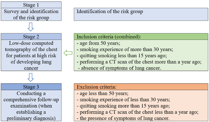
Lung cancer remains one of the most pressing medical and social problems of modern oncology, occupying a leading position in terms of morbidity and mortality in the world (Balditsyna et al., 2019; Thai et al., 2021; Harðardottir et al., 2022). The high mortality rate in this type of malignant neoplasm is mainly due to the late diagnosis of the disease, when the tumor reaches widespread stages and radical treatment becomes impossible (Alnemer et al., 2022; Deshpand et al., 2022; Kumar et al., 2022; Spirito et al., 2022; Prada & Ciavoi, 2024; Smolarz et al., 2025; Tbahriti et al., 2025). In this regard, a key task aimed at improving prognosis and reducing mortality is to detect lung cancer at early, asymptomatic stages, when therapy options are most effective (Nooreldeen & Bach, 2021; Lee & Kazerooni, 2022; AlHussain et al., 2023; Maneea et al., 2024). Figure 1 shows a diagram of the stages of lung screening.
|
|
|
Figure 1. Stages of lung cancer screening |
The notion of systematic screening of high-risk populations provided a solution to this dilemma (Saab et al., 2022). Large-scale randomized controlled trials, such as the National Lung Screening Trial (NLST) in the United States and the Dutch-Belgian NELSON study, have yielded convincing results: annual screening with low-dose computed tomography (LDCT) reduces lung cancer mortality by 20-24% when compared to chest X-ray (Oudkerk et al., 2021; Adams et al., 2023; Pan et al., 2023; Zhong et al., 2024). These findings served as the foundation for the integration of LDCT screening programs into clinical recommendations and real-world healthcare practice in a number of countries (Mazzone et al., 2021; Abdelmuhsin et al., 2022; Fiodorova et al., 2022; Lam et al., 2023; Zakinyan et al., 2023; Lam et al., 2024; Negreiros et al., 2024; Wolf et al., 2024).
However, significant practical and economic obstacles have arisen in the way of widespread implementation of screening. The central problem was the high detection rate of indeterminate pulmonary nodules, the vast majority of which subsequently turn out to be benign (Dickson et al., 2022; Lancaster et al., 2022; Masquelin et al., 2025). According to NLST data, the proportion of false positive results in the first round of screening reached 96.4% (Mao et al., 2025). A false positive result is defined as the detection of a CT scan of a tumor that is interpreted as suspicious of malignancy and requires additional examination but is not confirmed as cancer during a certain follow-up period (most often 1 year) (Kuś et al., 2014; Hammer et al., 2022; Lancaster et al., 2022).
The occurrence of false positive results entails a cascade of negative consequences:
Thus, one of the main tasks of a modern screening program is to find a balance between maximum sensitivity (the ability to identify all truly positive cases of cancer) and high specificity (the ability to avoid false alarms). Traditionally, this balance depends on the qualifications and experience of a radiologist, who must visually analyze huge amounts of data (hundreds and thousands of slices per study), which is a laborious process prone to fatigue, subjectivity, and variability of interpretation (Kates et al., 2021; Van De Luecht & Reed, 2021; Li et al., 2025).
The development of artificial intelligence (AI) technologies, in particular deep learning methods for image analysis, has opened a new era in medical imaging (Li et al., 2022; Huang et al., 2023). Computer vision systems trained on extensive datasets with annotations have demonstrated outstanding abilities in solving problems of object detection, segmentation, and classification (Li et al., 2022; Chen et al., 2023). In radiology, AI algorithms have been used to create decision support systems (DSS) designed to assist the doctor (Hosni et al., 2020).
In relation to lung cancer screening for LDCT, AI-DSS solves several key tasks:
Theoretically, the introduction of AI should not only increase the sensitivity of screening by reducing the number of missed cancers but also increase its specificity, making it possible to more confidently characterize nodules as benign and thereby reduce the number of false positive results (Chassagnon et al., 2023; Quanyang et al., 2024).
Despite the rapid development of commercial and academic AI solutions, their integration into the real clinical workflow requires careful validation (Ansari et al., 2022; Ansari et al., 2024; Samyuktha & Syam, 2024; Suchy & Jurkowski, 2024). The question of how exactly an AI assistant affects the work of a radiologist in the context of the sensitivity/specificity balance remains open. Will automatic detection lead to an increase in the number of LPR due to overdiagnosis of minor findings? Or, on the contrary, will accurate quantification and standardization of the approach reduce subjective variability and reduce the cascade of unnecessary follow-up studies?
Validating the algorithms themselves against expert judgment or histology is the main focus of the majority of current research (Zhang & Chen, 2022; de Margerie-Mellon & Chassagnon, 2023). In actuality, though, AI collaborates with doctors rather than replacing them. Because of this, it is crucial to compare the efficacy of the "doctor + AI" combination to the solo work of a physician.
The goal of this study is to compare the diagnostic efficacy and frequency of false positive outcomes in a radiologist's independent interpretation of lung LDCT with his work utilizing an AI-based decision support system.
The findings of this study will be extremely useful in justifying the incorporation of AI technologies into routine screening programs, allowing us to predict their true impact on early lung cancer detection, as well as the healthcare economy and patients' psychological comfort (Rani et al., 2023; Ludovichetti et al., 2024).
Materials and Methods
A retrospective cross-sectional investigation was carried out using an examination of the LDCT research database. The local Ethics Committee of the Medical Faculty of Ingush State University accepted the study (Protocol No. EC-45/2023). Due to the retroactive nature of the work and the complete depersonalization of data, informed permission was not required.
The study included LDCT studies of 100 patients who met the criteria for lung cancer screening (age 50-80 years, smoking index ≥ 30 packs/year), performed between January 2022 and June 2025 at the Republican Oncological Dispensary named after G.M. Vedzizhev (Republic of Ingushetia, Russia).
Inclusion criteria: the presence of an LDCT study performed on a Siemens Somatom go.Now CT scanner, the presence of at least one lung nodule measuring 4-30 mm, and the presence of a verified final diagnosis. Exclusion criteria: pronounced examination artifacts, tumors larger than 30 mm, absence of histological verification or 24-month X-ray follow-up.
The final sample included 35 studies with malignant nodules (histologically verified by adenocarcinoma or squamous cell carcinoma), 45 studies with benign nodules (stable for ≥24 months), and 20 studies without significant nodules.
All the studies were performed on a Siemens Somatom go CT scanner. Now, according to the standard low-dose CT protocol: voltage 100 kV, current with automatic modulation (Care Dose 4D, range 30-50 Wt), slice thickness 1.0 mm with reconstruction by the Br40f algorithm, and estimated effective radiation dose 1.2 ± 0.3 mSv.
The Philips IntelliSpace Discovery 3.0 artificial intelligence decision support system with the Lung Texture Analysis module was used. The deep learning-based algorithm provides automatic detection, segmentation, and quantitative characterization of pulmonary nodules, including calculation of volume parameters and automatic categorization according to the Lung-RADS scale version 2022.
Four radiologists participated in the study: one expert with more than 15 years of experience (MD, Professor of the Department of Radiation Diagnostics) and three second- and third-year residents. The assessment was conducted in two rounds with a cooling period of 8 weeks. In the first round, all radiologists independently analyzed the studies in the Sectra PACS software version 22.1 without access to the results of AI analysis. For each case, the presence and characteristics of suspicious nodules, the Lung-RADS category, recommendations for further tactics, and the time of analysis were recorded. In the second round, after a cooling-off period, the same radiologists reanalyzed the studies in random order with access to the AI analysis results displayed as overlay markings in the PACS interface. The same parameters were recorded as in the first round.
The final diagnosis was established on the basis of histological verification for malignant nodules (biopsy under CT navigation or material after surgery) (Patatou et al., 2022; Chen et al., 2024; Seoane-Viaño et al., 2024). For benign nodules, the criterion of absence of growth for 24 months was used according to dynamic LDCT observation data. The conclusions for the group without significant nodules were verified by a consensus decision of two independent experts who did not participate in the main study.
The primary endpoints were sensitivity, specificity, and frequency of false positive results. McNemar's test for paired proportions was used to compare diagnostic parameters between rounds. Interoperational consistency was assessed using the Kappa-Fleiss coefficient (Gwet, 2021). The time cost comparison was performed using the Student's paired t-test. The statistical analysis was performed in the IBM SPSS Statistics 29.0 and R 4.2.1 software environments. The statistical significance level was set to p < 0.05.
Results and Discussion
The analysis of diagnostic effectiveness was carried out on the basis of 100 studies, which included 80 nodules (35 malignant and 45 benign) and 20 cases without significant changes. The indicators of four radiologists were compared: an expert and three residents with an independent assessment and with the use of an AI assistant. Table 1 shows a comparison of diagnostic parameters of radiologists without AI and with an AI assistant. Table 2 shows the time required to analyze a single study. Tables 3 and 4 contain data on the distribution of Lung-RADS categories for false positive results and interoperable consistency.
Table 1. Comparison of diagnostic parameters of radiologists without AI and with an AI assistant
|
Indicator |
Group |
Without AI (%) |
With AI (%) |
p- value |
|
Sensitivity |
Expert |
94.3 |
97.1 |
0.317 |
|
Resident 1 |
80.0 |
91.4 |
0.021 |
|
|
Resident 2 |
77.1 |
88.6 |
0.039 |
|
|
Resident 3 |
74.3 |
85.7 |
0.046 |
|
|
Specificity |
Expert |
92.3 |
93.8 |
0.564 |
|
Resident 1 |
83.1 |
89.2 |
0.033 |
|
|
Resident 2 |
80.0 |
86.2 |
0.041 |
|
|
Resident 3 |
78.5 |
84.6 |
0.048 |
|
|
Frequency of false positive results |
Expert |
7.7 |
6.2 |
0.564 |
|
Resident 1 |
16.9 |
10.8 |
0.033 |
|
|
Resident 2 |
20.0 |
13.8 |
0.041 |
|
|
Resident 3 |
21.5 |
15.4 |
0.048 |
Table 2. Time spent on the analysis of one study (seconds)
|
Group |
Without AI (M ± SD) |
With AI (M ± SD) |
p-value |
|
Expert |
128 ± 23 |
94 ± 18 |
<0.001 |
|
Resident 1 |
215 ± 34 |
142 ± 27 |
<0.001 |
|
Resident 2 |
238 ± 41 |
156 ± 32 |
<0.001 |
|
Resident 3 |
251 ± 39 |
163 ± 29 |
<0.001 |
Table 3. Distribution of Lung-RADS categories for false positive results
|
Category |
Without AI (n=52) |
With AI (n=36) |
Δ, % |
|
2 |
8 (15.4%) |
12 (33.3%) |
+17.9 |
|
3 |
21 (40.4%) |
16 (44.4%) |
+4.0 |
|
4A |
15 (28.8%) |
6 (16.7%) |
-12.1 |
|
4B |
8 (15.4%) |
2 (5.6%) |
-9.8 |
Table 4. Inter-operator consistency (Fleiss' kappa)
|
Condition |
All doctors |
Residents |
Expert vs Residents |
|
Without AI |
0.62 |
0.58 |
0.71 |
|
With AI |
0.81 |
0.79 |
0.85 |
The study demonstrates the significant impact of the AI assistant system on the diagnostic parameters of radiologists in the analysis of lung LDCT. The most pronounced effect was observed among residents, which indicates the potential role of AI as a tool for standardization and training.
The increase in sensitivity by 11.4-14.3% in the group of residents (p<0.05) is consistent with the data from other studies and is explained by the AI's ability to detect small and low-contrast nodules, which are often overlooked by inexperienced specialists (Zhu et al., 2022; Mir et al., 2023; Weaver et al., 2025). It is important to note that AI did not show a significant improvement in the expert's performance (Δ+2.8%, p=0.317), which confirms the thesis about the ceiling effect for highly qualified specialists (Hunter et al., 2022; Esmaeilzadeh, 2024).
The decrease in the frequency of false positive results by 6.1-10.7% in the group of residents is of particular clinical interest. The analysis of the structure of false positive results revealed a significant reduction in categories 4A and 4B (Table 3), which directly affects the reduction in the number of unnecessary invasive procedures. This effect can be explained by an accurate quantitative assessment of the volume and density of nodules by the AI system, which reduces subjective overdiagnosis (Callister et al., 2021; Yankelevitz & Henschke, 2021; Xie et al., 2025).
Reducing the analysis time by 26.6-35.1% across all groups is of great practical importance for the implementation of screening programs. Saving 60-90 seconds on the study during mass screening can significantly increase the capacity of radiology departments (Hendrix et al., 2022).
The increase in the Kappa Fleiss coefficient from 0.62 to 0.81 indicates the important role of AI in standardizing the interpretation of research. The improvement in consistency between residents is particularly significant (from 0.58 to 0.79), which indicates the potential of AI as a tool to reduce interoperative variability (Sadoughi et al., 2022; Vanacore & Pellegrino, 2022; Robey et al., 2023).
Limitations of the study include a retrospective design and a relatively small sample size. Further prospective studies are required to assess the long-term impact of AI on clinical outcomes and the cost-effectiveness of screening programs.
The introduction of an AI assistant into the practice of lung LDCT analysis significantly improves the diagnostic performance of novice specialists, reduces the study time, and increases the standardization of diagnostics. The greatest benefit from the introduction of AI is expected in institutions with a high proportion of young professionals and a large volume of screening studies.
Conclusion
The study demonstrates the significant potential of integrating artificial intelligence systems into the clinical practice of analyzing low-dose computed tomography of the lungs. The results obtained indicate a comprehensive positive effect of the AI assistant on the diagnostic process. The most pronounced effect was observed in the group of residents, where the use of a decision support system increased sensitivity by 11.4-14.3% and reduced the frequency of false positive results by 6.1-10.7%. Of particular clinical importance is the reduction in the number of false positive conclusions of categories 4A and 4B on the Lung-RADS scale, which directly leads to a decrease in the number of unjustified invasive procedures.
An important practical result was a reduction in the analysis time of one study by 26.6-35.1% across all groups, which can significantly increase the capacity of radiology departments during mass screening examinations. The increase in the coefficient of interoperative consistency from 0.62 to 0.81 confirms the role of AI systems as a tool for standardizing diagnostic approaches and reducing subjective variability in the interpretation of results.
The information gathered allows for the recommendation that artificial intelligence technologies be incorporated into the standard clinical procedures of organizations carrying out screening initiatives for the early diagnosis of lung cancer. In medical organizations with a large number of young professionals and a substantial quantity of screening research, the employment of an AI assistant can be most useful. The evaluation of the cost-effectiveness of screening programs and the long-term effects of AI deployment on patient clinical outcomes, as well as the creation of adaptive learning systems with AI technology, are promising areas for more study.
Acknowledgments: The authors would like to thank all participants involved in this study. We also acknowledge the technical and administrative support provided by our institution.
Conflict of interest: None
Financial support: None
Ethics statement: The study was conducted in accordance with ethical standards and approved by the relevant institutional review board. Informed consent was obtained from all participants where applicable.
Abdelmuhsin, A. A., Alghamdi, A. A., & Ibrahim, N. A. (2022). Evaluating the phenotypic and genotypic diversity of Plantago ciliata in the Ha’il region, Saudi Arabia. International Journal of Veterinary Research & Allied Sciences, 2(1), 15–23. doi:10.51847/Qd2C6vFTgc
Adams, S. J., Stone, E., Baldwin, D. R., Vliegenthart, R., Lee, P., & Fintelmann, F. J. (2023). Lung cancer screening. Lancet, 401(10374), 390–408. doi:10.1016/S0140-6736(22)01694-4
AlHussain, B. S., AlShehri, A. M., AlRasheed, M. A., AlGadhi, S. K., & AlAhmad, F. A. (2023). A systematic review on endodontic retreatment and the effective removal of endodontic sealers using lasers. International Journal of Dental Research & Allied Sciences, 3(1), 8–16. doi:10.51847/m7WmmgNWH1
Alnemer, S., Alajlan, A. M., Alqarni, A. N., Alshanbari, S. H., Alhejazi, M. A., Matrood, M. A., Alkathiri, M. S., Almutairi, M. S., Aldayhani, A. B., Daabash, M. D., et al. (2022). Knowledge and practices of Riyadh-based dentists in managing traumatic dental injuries. Annals of Journal of Dental & Medical Assistance, 2(1), 22–25. doi:10.51847/ZGZXIiiSUR
Ansari, S. H., Qamar, Z., Alshammari, M., Bazoun, R., Alenazi, R., & Alattar, R. (2024). Clinical efficacy and longevity of monolithic vs. layered zirconia crowns; A systematic review. Bulletin of Pioneer Research in Medical & Clinical Sciences, 3(1), 7–18. doi:10.51847/0xBNKCjrDi
Ansari, S., Alshamrani, B., Alzahrani, A., Alfayez, A., Alhebshan, N., & Alshamrani, A. (2022). Prevalence of dental fluorosis among teenagers; A cross-sectional study in the schools of Riyadh. Bulletin of Pioneer Research in Medical & Clinical Sciences, 1(1), 13–17. doi:10.51847/37FuXGVEpm
Aresta, G., Ferreira, C., Pedrosa, J., Araujo, T., Rebelo, J., Negrao, E., Morgado, M., Alves, F., Cunha, A., Ramos, I., et al. (2020). Automatic lung nodule detection combined with gaze information improves radiologists' screening performance. IEEE Journal of Biomedical and Health Informatics, 24(10), 2894–2901. doi:10.1109/JBHI.2020.2976150
Attenborough, J., Abbott, S., Brook, J., & Knight, R. (2023). Studying barriers to work-based learning in clinical environments from the perspective of nursing managers and nurses. Journal of Integrated Nursing & Palliative Care, 4, 46–52. doi:10.51847/qQR0GNUES7
Balditsyna, E., Baklanov, I., & Baklanova, O. (2019). Gender identity studies in the modern social theory: research features. WinRS, 2, 43–51. doi:10.21064/winrs.2019.2.4
Bonney, A., Malouf, R., Marchal, C., Manners, D., Fong, K. M., Marshall, H. M., Irving, L. B., & Manser, R. (2022). Impact of low-dose computed tomography (LDCT) screening on lung cancer-related mortality. Cochrane Database of Systematic Reviews, 8(8), CD013829. doi:10.1002/14651858.CD013829.pub2
Bradley, S. H., Bhartia, B. S., Callister, M. E., Hamilton, W. T., Hatton, N. L. F., Kennedy, M. P., Mounce, L. T., Shinkins, B., Wheatstone, P., & Neal, R. D. (2021). Chest X-ray sensitivity and lung cancer outcomes: a retrospective observational study. British Journal of General Practice, 71(712), e862–e868. doi:10.3399/BJGP.2020.1099
Callister, M. E. J., Sasieni, P., & Robbins, H. A. (2021). Overdiagnosis in lung cancer screening. The Lancet Respiratory Medicine, 9(1), 7–9. doi:10.1016/S2213-2600(20)30553-1
Chassagnon, G., De Margerie-Mellon, C., Vakalopoulou, M., Marini, R., Hoang-Thi, T. N., Revel, M. P., & Soyer, P. (2023). Artificial intelligence in lung cancer: current applications and perspectives. Japanese Journal of Radiology, 41(3), 235–244. doi:10.1007/s11604-022-01359-x
Chen, M., Copley, S. J., Viola, P., Lu, H., & Aboagye, E. O. (2023). Radiomics and artificial intelligence for precision medicine in lung cancer treatment. Seminars in Cancer Biology, 93, 97–113. doi:10.1016/j.semcancer.2023.05.004
Chen, Z., Meng, L., Xiao, Y., Zhang, J., Zhang, X., Wei, Y., He, X., Zhang, X., & Zhang, X. (2024). Clinical application of optical and electromagnetic navigation system in CT-guided radiofrequency ablation of lung metastases. International Journal of Hyperthermia, 41(1), 2300333. doi:10.1080/02656736.2023.2300333
Cirik, V. A., Aksoy, B., & Bulut, E. (2023). Studying the relationship between the attitude towards gender roles of parents and the quality of parent-child relationship in nurses. Journal of Integrated Nursing & Palliative Care, 4, 30–37. doi:10.51847/90pkztCQgl
Damhus, C. S., Quentin, J. G., Malmqvist, J., Siersma, V., & Brodersen, J. (2021). Psychosocial consequences of a three-month follow-up after receiving an abnormal lung cancer CT-screening result: a longitudinal survey. Lung Cancer, 155, 46–52. doi:10.1016/j.lungcan.2021.03.003
de Margerie-Mellon, C., & Chassagnon, G. (2023). Artificial intelligence: a critical review of applications for lung nodule and lung cancer. Diagnostic and Interventional Imaging, 104(1), 11–17. doi:10.1016/j.diii.2022.11.007
Deshpand, R., Chandra, M., & Rauthan, A. (2022). Evolving trends in lung cancer: epidemiology, diagnosis, and management. Indian Journal of Cancer, 59(Supplement), S90–S105. doi:10.4103/ijc.IJC_52_21
Dickson, J. L., Bhamani, A., Quaife, S. L., Horst, C., Tisi, S., Hall, H., Verghese, P., Creamer, A., Prendecki, R., McCabe, J., et al. (2022). The reporting of pulmonary nodule results by letter in a lung cancer screening setting. Lung Cancer, 168, 46–49. doi:10.1016/j.lungcan.2022.04.009
Esmaeilzadeh, P. (2024). Challenges and strategies for wide-scale artificial intelligence (AI) deployment in healthcare practices: a perspective for healthcare organizations. Artificial Intelligence in Medicine, 151, 102861. doi:10.1016/j.artmed.2024.102861
Fay, M. P., & Lumbard, K. (2021). Confidence intervals for difference in proportions for matched pairs compatible with exact McNemar's or sign tests. Statistics in Medicine, 40(5), 1147–1159. doi:10.1002/sim.8829
Fiodorova, O. A., Sivkova, E. I., & Nikonov, A. A. (2022). Safeguarding beef cattle from gnats and gadflies in the southern Tyumen region. International Journal of Veterinary Research & Allied Sciences, 2(2), 8–13. doi:10.51847/iVXOeXmSNZ
Gwet, K. L. (2021). Large-sample variance of Fleiss generalized kappa. Educational and Psychological Measurement, 81(4), 781–790. doi:10.1177/0013164420973080
Hammer, M. M., Byrne, S. C., & Kong, C. Y. (2022). Factors influencing the false positive rate in CT lung cancer screening. Academic Radiology, 29(Suppl 2), S18–S22. doi:10.1016/j.acra.2020.07.040
Harðardottir, H., Jonsson, S., Gunnarsson, O., Hilmarsdottir, B., Asmundsson, J., Gudmundsdottir, I., Saevarsdottir, V. Y., Hansdottir, S., Hannesson, P., & Gudbjartsson, T. (2022). Advances in lung cancer diagnosis and treatment - a review. Laeknabladid, 108(1), 17–29. doi:10.17992/lbl.2022.01.671
Hendrix, N., Veenstra, D. L., Cheng, M., Anderson, N. C., & Verguet, S. (2022). Assessing the economic value of clinical artificial intelligence: challenges and opportunities. Value in Health, 25(3), 331–339. doi:10.1016/j.jval.2021.08.015
Hosni, M., García-Mateos, G., Carrillo-de-Gea, J. M., Idri, A., & Fernández-Alemán, J. L. (2020). A mapping study of ensemble classification methods in lung cancer decision support systems. Medical & Biological Engineering & Computing, 58(10), 2177–2193. doi:10.1007/s11517-020-02223-8
Huang, S., Yang, J., Shen, N., Xu, Q., & Zhao, Q. (2023). Artificial intelligence in lung cancer diagnosis and prognosis: current application and future perspective. Seminars in Cancer Biology, 89, 30–37. doi:10.1016/j.semcancer.2023.01.006
Hunter, B., Hindocha, S., & Lee, R. W. (2022). The role of artificial intelligence in early cancer diagnosis. Cancers (Basel), 14(6), 1524. doi:10.3390/cancers14061524
İlhan, N., Telli, S., Temel, B., & Aştı, T. (2022). Investigating the sexual satisfaction mediating role in the relationship between health literacy and self-care of men with diabetes and women's marital satisfaction. Journal of Integrated Nursing & Palliative Care, 3, 19–25. doi:10.51847/sFjL3OLpqg
Kates, F. R., Romero, R., Jones, D., Egelfeld, J., & Datta, S. (2021). A comparison of web-based cancer risk calculators that inform shared decision-making for lung cancer screening. Journal of General Internal Medicine, 36(6), 1543–1552. doi:10.1007/s11606-021-06754-0
Kumar, D., Gurunathan, D., Jabin, Z., & Talal, S. (2022). Comparative efficacy of aromatherapy and conscious sedation in pediatric dental anxiety management. Annals of Journal of Dental & Medical Assistance, 2(1), 14–21. doi:10.51847/TVRFhVaiIQ
Kuś, E., Gabryś, J., & Czyżewski, D. (2014). False positive result of 18F-FDG PET in patient with lung cancer due to sarcoid-like reaction in regional lymph nodes. Pneumonologia i Alergologia Polska, 82(6), 548–554. doi:10.5603/PiAP.2014.0072
Lam, D. C., Liam, C. K., Andarini, S., Park, S., Tan, D. S. W., Singh, N., Jang, S. H., Vardhanabhuti, V., Ramos, A. B., Nakayama, T., et al. (2023). Lung cancer screening in Asia: an expert consensus report. Journal of Thoracic Oncology, 18(10), 1303–1322. doi:10.1016/j.jtho.2023.06.014
Lam, S., Bai, C., Baldwin, D. R., Chen, Y., Connolly, C., de Koning, H., Heuvelmans, M. A., Hu, P., Kazerooni, E. A., Lancaster, H. L., et al. (2024). Current and future perspectives on computed tomography screening for lung cancer: a roadmap from 2023 to 2027 from the International Association for the Study of Lung Cancer. Journal of Thoracic Oncology, 19(1), 36–51. doi:10.1016/j.jtho.2023.07.019
Lancaster, H. L., Heuvelmans, M. A., & Oudkerk, M. (2022). Low-dose computed tomography lung cancer screening: clinical evidence and implementation research. Journal of Internal Medicine, 292(1), 68–80. doi:10.1111/joim.13480
Lancaster, H. L., Zheng, S., Aleshina, O. O., Yu, D., Yu Chernina, V., Heuvelmans, M. A., de Bock, G. H., Dorrius, M. D., Gratama, J. W., Morozov, S. P., et al. (2022). Outstanding negative prediction performance of solid pulmonary nodule volume AI for ultra-LDCT baseline lung cancer screening risk stratification. Lung Cancer, 165, 133–140. doi:10.1016/j.lungcan.2022.01.002
Lee, E., & Kazerooni, E. A. (2022). Lung cancer screening. Seminars in Respiratory and Critical Care Medicine, 43(6), 839–850. doi:10.1055/s-0042-1757885
Li, Y., Law, J., Le, L. W., Li, J. J. N., Pettengell, C., Demarco, P., Duong, M., Merritt, D., Davidson, S., Sung, M., et al. (2025). Assessing the feasibility and external validity of natural language processing-extracted data for advanced lung cancer patients. Lung Cancer, 199, 108080. doi:10.1016/j.lungcan.2025.108080
Li, Y., Wu, X., Yang, P., Jiang, G., & Luo, Y. (2022). Machine learning for lung cancer diagnosis, treatment, and prognosis. Genomics, Proteomics & Bioinformatics, 20(5), 850–866. doi:10.1016/j.gpb.2022.11.003
Ludovichetti, F. S., Stellini, E., Zuccon, A., Lucchi, P., Dessupoiu, N., Mazzoleni, S., & Parcianello, R. G. (2024). Comparative impact of chlorhexidine and fluoride varnish on white spot lesion prevention in orthodontic patients. Turkish Journal of Public Health & Dentistry, 4(1), 6–12. doi:10.51847/Pw5eiJPGY2
Maneea, A. S. B., Alqahtani, A. D., Alhazzaa, A. K., Albalawi, A. O., Alotaibi, A. K., & Alanazi, T. F. (2024). Systematic review of the microbiological impact of sodium hypochlorite concentrations in endodontic treatment. International Journal of Dental Research & Allied Sciences, 4(2), 9–15. doi:10.51847/PH80PpWOX7
Mao, Y., Lancaster, H. L., Heuvelmans, M. A., Han, D., Yu, D., Yi, J., Gratama, J. W. C., de Bock, G. H., Oudkerk, M., Ye, Z., et al. (2025). Comparison of nodule volumetric classification by using two different nodule segmentation algorithms in an LDCT lung cancer baseline screening dataset. European Journal of Radiology, 191, 112290. doi:10.1016/j.ejrad.2025.112290
Masquelin, A. H., Cheney, N., José Estépar, R. S., Bates, J. H. T., & Kinsey, C. M. (2025). LDCT image biomarkers that matter most for the deep learning classification of indeterminate pulmonary nodules. Cancer Biomarkers, 42(1), CBM230444. doi:10.3233/CBM-230444
Mazzone, P. J., Silvestri, G. A., Souter, L. H., Caverly, T. J., Kanne, J. P., Katki, H. A., Wiener, R. S., & Detterbeck, F. C. (2021). Screening for lung cancer: CHEST guideline and expert panel report. Chest, 160(5), e427–e494. doi:10.1016/j.chest.2021.06.063
Michael, C. W., & Engels, M. (2025). Benign category in the WHO reporting system for lung cytopathology: diagnostic difficulties and differential diagnosis. Cytopathology, 36(5), 443–457. doi:10.1111/cyt.13497
Mir, M. M., Mir, G. M., Raina, N. T., Mir, S. M., Mir, S. M., Miskeen, E., Alharthi, M. H., & Alamri, M. M. S. (2023). Application of artificial intelligence in medical education: current scenario and future perspectives. Journal of Advanced Medical Education & Professionalism, 11(3), 133–140. doi:10.30476/JAMP.2023.98655.1803
Mobeen, T., & Dawood, S. (2022). Studying the effect of perceived social support and mental health on marital burnout in infertile women. Journal of Integrated Nursing & Palliative Care, 3, 7–12. doi:10.51847/7DkM3Fkiu3
Morris, M. J., Habib, S. A., Do Valle, M. L., & Schneider, J. E. (2024). Economic evaluation of a novel lung cancer diagnostic in a population of patients with a positive low-dose computed tomography result. Journal of Health Economics and Outcomes Research, 11(2), 74–79. doi:10.36469/001c.121512
Negreiros, A. B., Silva, G. R. D., Pereira, F. D. M., Souza, B. D. A., Lopes, M. T. D. R., & Diniz, F. M. (2024). Evidence of genetic diversity gradients in Melipona rufiventris (Hymenoptera: Apidae) within the Brazilian semiarid region. Entomology Letters, 4(1), 1–7. doi:10.51847/l9Wmr8r6qW
Nooreldeen, R., & Bach, H. (2021). Current and future development in lung cancer diagnosis. International Journal of Molecular Sciences, 22(16), 8661. doi:10.3390/ijms22168661
Oudkerk, M., Liu, S., Heuvelmans, M. A., Walter, J. E., & Field, J. K. (2021). Lung cancer LDCT screening and mortality reduction - evidence, pitfalls and future perspectives. Nature Reviews Clinical Oncology, 18(3), 135–151. doi:10.1038/s41571-020-00432-6
Pan, Z., Zhang, R., Shen, S., Lin, Y., Zhang, L., Wang, X., Ye, Q., Wang, X., Chen, J., Zhao, Y., et al. (2023). OWL: an optimized and independently validated machine learning prediction model for lung cancer screening based on the UK Biobank, PLCO, and NLST populations. EBioMedicine, 88, 104443. doi:10.1016/j.ebiom.2023.104443
Patatou, A., Iacovou, N., Zaxaria, P., Vasoglou, M., & Vasoglou, G. (2022). Corticotomy-assisted orthodontics: biological basis and clinical applications. Annals of Orthodontics & Periodontics Special, 2, 8–13. doi:10.51847/0qGERVSoQm
Pei, Q., Luo, Y., Chen, Y., Li, J., Xie, D., & Ye, T. (2022). Artificial intelligence in clinical applications for lung cancer: diagnosis, treatment and prognosis. Clinical Chemistry and Laboratory Medicine, 60(12), 1974–1983. doi:10.1515/cclm-2022-0291
Prada, A. M., Cicalău, G. I. P., & Ciavoi, G. (2024). Resin infiltration for white-spot lesion management after orthodontic treatment. Asian Journal of Periodontics & Orthodontics, 4, 19–23. doi:10.51847/ZTuGEanCSV
Quanyang, W., Yao, H., Sicong, W., Linlin, Q., Zewei, Z., Donghui, H., Hongjia, L., & Shijun, Z. (2024). Artificial intelligence in lung cancer screening: detection, classification, prediction, and prognosis. Cancer Medicine, 13(7), e7140. doi:10.1002/cam4.7140
Rani, H., Mohd-Dom, T. N., Meei, T. I., Rosli, M. S., Quan, L. Z., Aziz, A. F., Hassan, S. A., & Aun, N. S. (2023). Dental home care challenges for homebound patients at Prince Sultan Military Medical City. Turkish Journal of Public Health & Dentistry, 3(2), 27–32. doi:10.51847/VwQFNrdHml
Robey, C., McCammon, K., Perry, M., Zagade, T., Brugh, V., & Liu, J. (2023). Inter-rater reliability of retrograde urethrograms. World Journal of Urology, 41(4), 1163–1167. doi:10.1007/s00345-023-04323-0
Saab, M. M., O'Driscoll, M., Sahm, L. J., Leahy-Warren, P., Noonan, B., FitzGerald, S., Kilty, C., O'Malley, M., Lyons, N., & Hegarty, J. (2022). Referring high-risk individuals for lung cancer screening: a systematic review of interventions with healthcare professionals. European Journal of Cancer Prevention, 31(6), 540–550. doi:10.1097/CEJ.0000000000000755
Sadoughi, F., Behmanesh, A., Najd Mazhar, F., Joghataei, M. T., Yazdani, S., Shams, R., Morovvati, H., Najaf Asaadi, S., & Vosough, A. (2022). Bone healing monitoring in bone lengthening using bioimpedance. Journal of Healthcare Engineering, 2022, 3226440. doi:10.1155/2022/3226440
Samyuktha, P. S., & Syam, S. (2024). Periodontal abscess as a clinical oral sign in patients with diabetes mellitus - An original study. Bulletin of Pioneer Research in Medical & Clinical Sciences, 3(2), 7–12. doi:10.51847/ZDpdihizWm
Seoane-Viaño, I., Seoane-Gigirey, M., Bendicho-Lavilla, C., Gigirey, L. M., Otero-Espinar, F. J., & Seoane-Trigo, S. (2024). Localized drug delivery using linezolid hydrogel for periodontal therapy. Annals of Orthodontics & Periodontics Special, 4, 39–46. doi:10.51847/nSySWrEvcx
Siwik, C. J., Phillips, K., Zimmaro, L., Salmon, P., & Sephton, S. E. (2022). Depressive symptoms among patients with lung cancer: elucidating the roles of shame, guilt, and self-compassion. Journal of Health Psychology, 27(5), 1039–1047. doi:10.1177/1359105320988331
Smolarz, B., Łukasiewicz, H., Samulak, D., Piekarska, E., Kołaciński, R., & Romanowicz, H. (2025). Lung cancer-epidemiology, pathogenesis, treatment and molecular aspect (Review of literature). International Journal of Molecular Sciences, 26(5), 2049. doi:10.3390/ijms26052049
Spirito, F. D., Iacono, V. J., Alfredo, I., Alessandra, A., Sbordone, L., & Lanza, A. (2022). Impact of COVID-19 awareness on periodontal disease prevention and management. Asian Journal of Periodontics & Orthodontics, 2, 16–26. doi:10.51847/t8D9TJGOCU
Suchy, W., & Jurkowski, O. (2024). Clinical assessment of 5% lidocaine patches for postoperative analgesia: efficacy, effectiveness, and safety. Bulletin of Pioneer Research in Medical & Clinical Sciences, 3(1), 31–36. doi:10.51847/UxKg3AkOTB
Tbahriti, H. F., Zerrouki, A., Boukadoum, A., Kameche, M., Povetkin, S., Simonov, A., & Thiruvengadam, M. (2025). Comprehensive review and meta-analysis of magnesium chloride optimization in PCR: investigating concentration effects on reaction efficiency and template specificity. Analytical Biochemistry, 705, 115909. doi:10.1016/j.ab.2025.115909
Ten Haaf, K., van der Aalst, C. M., de Koning, H. J., Kaaks, R., & Tammemägi, M. C. (2021). Personalising lung cancer screening: an overview of risk-stratification opportunities and challenges. International Journal of Cancer, 149(2), 250–263. doi:10.1002/ijc.33578
Thai, A. A., Solomon, B. J., Sequist, L. V., Gainor, J. F., & Heist, R. S. (2021). Lung cancer. Lancet, 398(10299), 535–554. doi:10.1016/S0140-6736(21)00312-3
Tian, X., Yi, L. J., Liang, C. S., Gu, L., Peng, C., Chen, G. H., & Jiménez-Herrera, M. F. (2022). The impact of mindfulness-based stress reduction (MBSR) on psychological outcomes and quality of life in patients with lung cancer: a meta-analysis. Frontiers in Psychology, 13, 901247. doi:10.3389/fpsyg.2022.901247
Van De Luecht, M. R., & Reed, W. M. (2021). The cognitive and perceptual processes that affect observer performance in lung cancer detection: a scoping review. Journal of Medical Radiation Sciences, 68(2), 175–185. doi:10.1002/jmrs.456
Vanacore, A., & Pellegrino, M. S. (2022). Robustness of κ-type coefficients for clinical agreement. Statistics in Medicine, 41(11), 1986–2004. doi:10.1002/sim.9341
Vergnenegre, A., & Chouaid, C. (2021). Economic analyses of immune-checkpoint inhibitors to treat lung cancer. Expert Review of Pharmacoeconomics & Outcomes Research, 21(3), 365–371. doi:10.1080/14737167.2021.1863790
Weaver, C., Caputo, N. J., Yost, C., & Hauert, S. A. (2025). Using an AI-Nurse to help teach graduating medical students to take overnight call. Medical Science Educator, 35(2), 615–617. doi:10.1007/s40670-025-02335-6
Wolf, A. M. D., Oeffinger, K. C., Shih, T. Y., Walter, L. C., Church, T. R., Fontham, E. T. H., Elkin, E. B., Etzioni, R. D., Guerra, C. E., Perkins, R. B., et al. (2024). Screening for lung cancer: 2023 guideline update from the American Cancer Society. CA: A Cancer Journal for Clinicians, 74(1), 50–81. doi:10.3322/caac.21811
Wong, C. W. T., Xuen Lee, J. Z., Jaeschke, A., Ng, S. S. Y., Lit, K. K., Wan, H. Y., Kniebs, C., Ker, D. F. E., Tuan, R. S., & Blocki, A. (2025). Lung cancer intravasation-on-a-chip: visualization and machine learning-assisted automatic quantification. Bioactive Materials, 51, 858–875. doi:10.1016/j.bioactmat.2025.06.028
Xie, D., Zhang, L., He, N., Yang, C., Zhang, R., Chen, H., Liu, X., Suo, C., Wang, M., Wei, Y., et al. (2025). Overdiagnosis of lung cancer due to the introduction of low-dose computed tomography in average-risk populations in the People's Republic of China. Journal of Thoracic Oncology, 20(7), 884–896. doi:10.1016/j.jtho.2025.02.013
Yankelevitz, D. F., & Henschke, C. I. (2021). Overdiagnosis in lung cancer screening. Translational Lung Cancer Research, 10(2), 1136–1140. doi:10.21037/tlcr-20-736
Zakinyan, R. G., Badakhova, G. K., Lopteva, M. S., Koshkina, N. A., Tolokonnikov, V. P., & Povetkin, S. N. (2023). The link between ixodid tick populations and climate change in the Stavropol region. Entomology Letters, 3(2), 38–43. doi:10.51847/4lXTZ8h0Bs
Zhang, K., & Chen, K. (2022). Artificial intelligence: opportunities in lung cancer. Current Opinion in Oncology, 34(1), 44–53. doi:10.1097/CCO.0000000000000796
Zhong, D., Sidorenkov, G., Jacobs, C., de Jong, P. A., Gietema, H. A., Stadhouders, R., Nackaerts, K., Aerts, J. G., Prokop, M., Groen, H. J. M., et al. (2024). Lung nodule management in low-dose CT screening for lung cancer: lessons from the NELSON trial. Radiology
Zhu, S., Gilbert, M., Chetty, I., & Siddiqui, F. (2022). The 2021 landscape of FDA-approved artificial intelligence/machine learning-enabled medical devices: an analysis of the characteristics and intended use. International Journal of Medical Informatics, 165, 104828. doi:10.1016/j.ijmedinf.2022.104828