
To compare the subfoveal, parafoveal, and peripapillary choroidal thickness in amblyopic eyes with fellow and control eyes. Twelve control eyes, six fellow eyes, and six amblyopic eyes in this cross-sectional study. Choroidal thickness was measured by the enhanced-depth imaging technique of optical coherence tomography in all subjects. Choroidal thickness was measured at the subfoveal area and at 1000 mm intervals to the nasal and temporal to the fovea up to 2000 mm. The mean age was 8.75 ± 2.73 years in controls and 9.50 ± 2.81 years in amblyopic patients. Parafoveal choroidal thickness was thickest subfoveally, but thinnest nasally, in all three groups. The mean subfoveal choroidal thickness was 256 ± 40 in control eyes, 226±44 in fellow eyes, and 270±34 in amblyopic eyes. It was thicker in amblyopic eyes but not significantly, P-value > 0.05. There was no significant result in choroidal thickness between amblyopic, fellow, and control eyes.
Introduction
Amblyopia
Amblyopia is a visual disorder where there is a decrease in visual acuity in one or both eyes (unilateral or bilateral) caused by binocular visual experience during the “critical period (Progress in Brain Research, 1963). There are three periods when visual acuity and ocular dominance are developed. In the 3-5 years, acuity is measured by tests that exclude any crowding effects from less than 20/200 to nearly 20/20 (Early Visual Development: Normal and Abnormal, 2019).
The critical period depends on the process being examined at the anatomical level. The retina extends into the lateral geniculate nucleus, which projects into the primary visual cortex, then into other regions of the occipital cortex, and eventually into the sensory areas of the parietal and temporal cortex (LeVay et al., 1980). Cells at the system's higher levels have a critical period that lasts longer. For example, monocular deprivation can change the ocular dominance of cells in layers II, III, V, and VI of the primary visual cortex after establishing the ocular dominance of cells in layer IV (LeVay et al., 1980; Toigawa et al., 2010). Monocular deprivation affects several visual functions, with critical periods occurring early for functions treated at lower system levels. For example, monocular deprivation in the macaque monkey affects absolute sensitivity to light before the age of 3 months, sensitivity to wavelength and light increases before the age of 6 months, sensitivity to high-frequency contrast before the age of 18 months, and binocular summation before the age of 24 months (Harwerth et al., 1986; Yawman, 2007).
Etiology
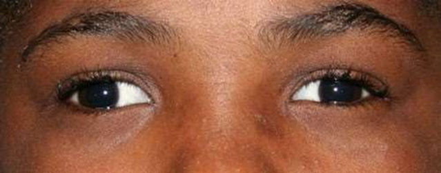
Amblyopia is a major problem in public health. In children and young adults, it is the most common cause of monocular vision loss (Williams, 2018). Amblyopia may be caused by several factors. Strabismus (ocular misalignment or heterotropia) causes the retinal images to be dissimilar. Uncorrected refractive errors (anisometropia) or visual axis obstructions (cataracts) it leads to unequal clarity of images. The most common contributing factor is strabismus. Deviating eye consistently leads to amblyopia, which may turn outward (exotropia) (Figure 1), inward (esotropia), or vertically (hypotropia, hypertropia). Deviations are detected in latent strabismus (heterophoria) only when one eye is occluded is rarely associated with amblyopia (Zhang & Wildsoet, 2015). The second most common factor contributing to amblyopia is `11``refractive error, which can be hard to detect.
Amblyopia is classified into two types arising from refractive errors: anisometropic and isometropic. Anisometropic amblyopia occurs when both eyes have different refractive errors, leading to the image being out of focus on one retina, impairing the visual pathway development in the affected eye. The most common refractive error that causes amblyopia is bilateral hyperopia (farsightedness). Isometropic amblyopia occurs when both eyes have the same refractive errors. Severe refractive errors can cause bilateral isometropic amblyopia in both eyes, even if symmetric, particularly if optical correction is delayed (Zhang & Wildsoet, 2015).
The least common factor is congenital opacity that causes amblyopia deprivation. Corneal lesions, ptosis, cataract, and distort the retinal image formation. Amblyopia of deprivation can affect one or both eyes and can take place within two to four months. Occlusion amblyopia is excessive therapeutic patching is a type of deprivation amblyopia (Zhang & Wildsoet, 2015).
|
|
|
Figure 1. child with exotropia of the right eye |
Diagnosis
Amblyopia is diagnosed when there is evidence of decreased visual acuity that physical anomalies cannot describe. Structural eye disorder should be excluded, including optic nerve or macular defects, media opacity, and central visual pathway abnormalities. The assessment starts by providing an eye history based on the patient’s age and parental observations (Zhang & Wildsoet, 2015). The parent or patient should be questioned about any obvious turns of the eye, the habit of closing one eye, depth difficulty, perceptual deficits, or school problems. The eye and visual examination should include a 1- monocular and binocular distance and near visual acuity using a whole chart, an isolated line, and an isolated letter to screen for crowding, 2- a dry and wet refraction to screen for latent hyperopia, and 3- analysis of monocular fixation to screen for eccentric fixation. 4- vergence, version, and sensorimotor fusion to screen for strabismus. 5- monocular and binocular oculomotor dysfunction testing motility. 6- accommodating amplitudes and accommodating dysfunction screening services, and 7- ocular safety, including color vision and dilated context screening for ocular disease or anatomical abnormalities that may result in reduced visual acuity (Zeiss et al., 2018).
Impact of Amblyopia
Previous studied the impact of amblyopia in children. The impact of amblyopia and the potential for reduced quality of life, the lifetime risk of bilateral vision impairment is almost double that for amblyopes than for controls, being as high as 18% for amblyopes compared to 10% for non-amblyopes (Corbett & Haines, 2018). Amblyopia does not have a significant impact on the outcomes of education, health, or society (Carreiro, 2009). Children with amblyopia do arithmetic, reading comprehension and visual and motor skills as well as their peers in age-appropriate assessments, and are usually less likely to have major behavioral problems or changes at home or school than those without amblyopia, while Adult amblyopes with moderate to severe visual acuity deficits (6/18 or worse) were more likely to report a road accident as the driver who caused hospital injuries (Spaide et al., 2008; Carreiro, 2009). Increased readability for children with amblyopia compared to those who have normal development in view is reported; nevertheless, the prevalence of specific reading disabilities is similar to that of the general population among amblyopia (Mrejen & Spaide, 2013). Amblyopic children have fewer motor skills than children with normal vision. These studies generally describe how amblyopia has an adverse effect on real-world visual-motor skills important for daily work, like reaching and capturing, drawing, writing, and manual skills (Dobson, 1993; Ikuno et al., 2011).
Prevalence
Worldwide, the prevalence of amblyopia is approximately 1%-5%. The World Health Organization (WHO) estimates that 19 million children under the age of 15 are visually impaired, 12 million of them are impaired due to uncorrected refractive errors and amblyopia (von Noorden, 1990). In different parts of the world, the prevalence of amblyopia varies,
with the highest incidence in Europe (Ahn et al., 2025; Castellano-Rioja, 2025; Cavero & Ferraz, 2025; Khalil & Nassar, 2025; Rani & Gehrke, 2025; Rivas & Molina, 2025; Shen & Bao, 2025; Silik, 2025). Geographical location and criteria for the definition of amblyopia are among the factors that contribute to the differences across the globe (Daw, 1998).
The prevalence of amblyopia in the US was less than (2%),
National population projections indicate that there are approximately 677,000 cases of manifest strabismus among children aged 6-71 months and 271,000 cases of amblyopia among children aged 30-71 months. The prevalence of amblyopia in Saudi Arabia were (1.85%) in Abha, anisometropic and strabismic amblyopia were (47%) and (13%) with deprivation amblyopia (LeVay et al., 1980), (3.90%) in Qassim, anisometropic amblyopia was (77.72%) and strabismic amblyopia was (5.44%) (Daw et al., 1992) and in Jeddah the prevalence was (1.3%), anisometropic was (69%) and strabismic was (6%) (Harwerth et al., 1986).
Treatment
Treatment is based on the cause of amblyopia and should be initiated at an early period (critical period). During this period, the various forms of deprivation listed above may reduce acuity, leading to amblyopia. Nevertheless, amblyopia is not limited to the first 3 to 5 years of life, but may result from strabismus or anisometropia at any age, between several months and 7 or 8 years of age (Levi & Polat, 1996). Therefore, three stages of acuity can be explained: the period of visual acuity development (birth to 3-5 years), the duration during which deprivation is significant to cause amblyopia (from a few months to 7 or 8 years of age), and the period during which amblyopia can be recovered (adolescent or even adult deprivation time) (LeVay et al., 1980). The retina develops into the lateral geniculate nucleus, which projects into the primary visual cortex, then into other parts of the occipital cortex, and eventually into the sensory regions of the parietal and temporal cortex. Cells at the system's higher levels have a critical period that lasts longer. For example, monocular deprivation may alter the ocular dominance of cells in layers II, III, V, and VI of the primary visual cortex after establishing the ocular dominance of cells in layer IV (Ing, 1983; von Noorden, 1990).
In relation to stereopsis, stereopsis will never be achieved unless amblyopia is treated, eyes are aligned, and binocular fusion and function are achieved before the end of the critical period for stereopsis. Clinical data suggested that this occurs before the age of 24 months (Jampolsky, 1978; Held & Birch, 1980; Birch & Hale, 1996). Stereoscopic acuity reaches between 2 and 5 months of age, 60 minutes of arc, and increases rapidly within the following month (Birch & Hale, 1996). There is a slower rise from 6 months to 3 years. It is expected that the stereopsis can be destroyed over a period of more than three years and recovered over an even longer period (Gregg & Parks, 1992; Ciner et al., 1996). In an extreme case of unilateral cataract, a long-term patching regimen on the day after birth induces 20/25 visual acuity of the treated eye in a person aged 8 years and 50 seconds of stereoacuity in the arc (Helveston, 1993). Also, intervening in congenital esotropia at about 4 months of age may have a good rationale (Wright et al., 1994), as this is the age at which stereopsis is rapidly developing. Two out of 7 patients treated at this age had stereoacuity greater than 60 seconds of arc at 5 to 8 years of age (American Academy of Ophthalmology, n.d.).
Detecting amblyopia, it depends on early recognition of factors, including a significant decrease in visual acuity. There are some screening tests to evaluate amblyopia: red reflex test, ocular motility, and visual assessment (Robaei et al., 2006). The treatment goal is to provide a focused retinal image with appropriate optical correction, which can be done by patching the better eye to enhance the brain's ability to use the blurred image. In patients with visual deprivation, the derivative factors should be removed medically or surgically. Spectacles are the first line of treatment in anisometropic amblyopia, or, less commonly, contact lenses. Cycloplegic agent atropine sulfate can be used to enhance the use of the amblyopic eye. A drop of atropine applied to the better eye will reduce the accommodative ability, will be reduced and the eye receives a blurred image (Rahi et al., 2006). Although atropine is just as effective as patches for the treatment of amblyopia, it is less controllable than patches because the effects of atropine last up to 2 weeks and rarely cause a decrease in the eye (Wilson & Welch, 2013). It is an acceptable form of treatment for certain children and their parents. This is known as reverse amblyopia. Therefore, every 2- 3 weeks, a child with an atropine penalty must be observed to monitor vision closely in both eyes. The risk of reversal amblyopia and the increased number of clinical visits are the main reasons why atropine penalization is not routinely used as the initial amblyopia treatment (Wilson & Welch, 2013).
As a practical consideration, most strabismologists agree that it is necessary to improve acuity in the amblyopic eye by patching or other means before aligning the eyes. Otherwise, after alignment, the patient will not maintain their eyes straight. There are many other sensory tasks after this procedure that can be treated by several orthoptic techniques. Such functions include single-letter acuity, binocular function, stereopsis, depth perception, sensitivity to contrast, position perception, vernier acuity, crowding phenomenon (acuity in line of letters close to each other) (Koklanis et al., 2006).
Also, the eye structures, such as the choroid, could be affected by amblyopia. Although the foveal or parafoveal choroidal thickness varies significantly regardless of the depth and type of amblyopia.
Choroid
The choroid is a vascular, thin, and loose connective tissue that lies between the retinal pigment epithelium (RPE) and sclera, which arises very early in development from the loose vascular layer of mesenchyme that surrounds the optic cup. The main function of the choroid is to provide a nourishing blood supply to the outer layers of the retina. The choroid is too pigmented so that it may absorb the light that has entered the eye and passed through the retina (O’Connor et al., 2010). Recent studies showed the choroidal role in ocular focus adjustment, including emmetropization, and thus eye growth regulation (Suttle et al., 2011). It has three layers: choriocapillaris, stroma, and lamina fusca (from inner to outer). Each layer has specific functions. The choriocapillaris is responsible for supplying the nutrients to the (RPE) and the outer third of the retina. The choroidal stroma includes dendritic melanocytes, fibroblasts, and mast cells. The lamina fusca serves as a thin, web-like attachment between the choroid and sclera. The choroidal vasculature is supplied by the long and short posterior ciliary arteries and the anterior ciliary arteries. Drainage occurs via the vortex vessels (Hashimoto et al., 2017). Cerebrospinal fluid (CSF) is produced from choroid epithelial cells by selective transport of materials from the connective tissue extracellular space and choroidal capillaries. Large molecules are transported in pinocytotic vesicles from the basal to the apical surface of the epithelium and exocytosed into the CSF (Hashimoto et al., 2017).
Chloride, magnesium, and sodium have a higher concentration in CSF, while potassium, calcium, glucose, proteins, albumin, and uric acid have lower concentrations (Corbett & Haines, 2018). The choroid is innervated by the long and short ciliary nerves, which pass through the choroid and transmit sensory fibers to the cornea, iris, and ciliary body, while
The short ciliary nerves originated from the ciliary ganglion and carry sensory (from nasociliary), sympathetic, and parasympathetic fibers (derived predominantly from nerve III, but also from VII). Amblyopia leads to changes in choroidal blood flow (CBF) and central choroidal thickness (CCT) (Brown & Regillo, 2007) and therefore will affect the retina by changing changes in retinal vessels density and retinal thickness, macular vessel density in the superficial capillary plexuses of foveal and parafoveal is lower in children with amblyopia, while the thicknesses of the inner and outer retinas at either the fovea or the parafovea showed no significant differences between the amblyopic and control children.
The choroid is affected by many diseases, including Vogt-Koyanagi-Harada syndrome (VKH), sarcoidosis, sympathetic ophthalmia, toxoplasmosis, Bechet’s disease, and panuveitis. As the choroid is placed under RPE, many medically available imaging tools, including the fundus fluorescein angiogram (FFA), the B-scan ultrasound, and the optical coherence tomography (OCT), only provide a partial description of the structure and function. This is primarily due to a loss of signal and light on the highly reflective RPE surface that blocks most choroidal signals (LeVay et al., 1980).
Optical Coherence Tomography (OCT)
To create cross-sectional images, OCT uses the principle of low-coherence interferometry to obtain in-depth information from different retinal structures. It is an extremely useful tool for imagining and identifying different retinal layers and helps identify retinal pathology. However, OCT cannot penetrate the retinal pigment epithelium (RPE), and it is limited to imaging the retina and optic nerve head.
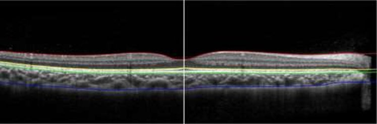
Enhanced depth imaging (EDI) enables imaging of the deeper structures, including the choroid. It provides detailed choroid information by moving the zero-delay point, the point of maximum sensitivity to the OCT signal. Placing the zero delay point closer to the choroid than the internal retinal layers leads to a better representation of the choroid and allows for high accuracy and reproducibility quantitative measurement of its thickness (Figure 2) (Daw et al., 1992; Friedman et al., 2009).
OCT imaging is widely used for macula, optic nerve, and retinal nerve fiber layer (RNFL) imaging and helps analyze the morphology and quantify changes in different disease states. For example, the automated retinal thickness measurements produced by the SD-OCT systems are medically used to track disease progressions such as wet age-related macular degeneration (AMD) and macular edema from various causes, including diabetes and occlusion of retinal veins. The ability to detect fluid in the retina and the thickness changes caused by fluid help guide medical decisions about treatment (Aldebasi, 2015).
|
|
|
Figure 2. Shows the choroidal and retinal layers. Choroidal thickness measured from RPE (green) to choroidoscleral interface (CSI) (blue). The center of the fovea is determined manually as the thinnest part of the retina (white). |
|
|
|
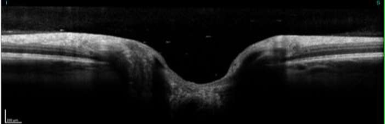
Figure 3. heathy optic nerve |
Literature Review
Fujiwara et al. in (2012) measured subfoveal choroidal thickness in normal Japanese eyes and found that subfoveal choroidal thickness was affected by age and refractive error; it was decreased in thickness every 10 years by 20 μm (Fujiwara et al., 2012). Another study by Bidaut et al. in (2013) measured the choroidal thickness in healthy children, and they found that the choroidal thickness was increased with age. Jinling et al. in (2014) Ebru et al. Ozgur et al. in (2015), and Zha et al. in (2019), used spectral-domain OCT to measure the choroidal thickness, subfoveal choroids in amblyopia subjects and found significantly thickening in both anisometropic and strabismic amblyopic eyes than those of the corresponding group of fellow eyes and control eyes.
In 2015, Feride et al. did a prospective study to evaluate the choroidal thickness and peripapillary retinal nerve fiber layer (RNFL) thickness in adults' amblyopic eyes. The choroidal thickness was thicker in the amblyopic eye than in the fellow eye, and there were no significant differences in RNFL thickness between amblyopic and fellow eyes (Kantarci et al., 2015). Another study in 2017 by Arzu et al. to compare the retina and optic disc between amblyopic and non-amblyopic eyes. RNFL thickness, macular thickness, macular volume, axial length, or optic disc area between amblyopic and non-amblyopic eyes showed no significant differences (Taşkıran Çömez et al., 2017). In 2017, Kaderli et al. did a study of the correlation of hyperopia and choroidal thickness, vessel diameter, and area, and they found that the macular choroidal thickness was thicker in hyperopic patients than in controls, so the choroidal thickness was affected by refractive error (Kaderli et al., 2017).
Materials and Methods
Aim of Study
To compare the subfoveal, parafoveal, and peripapillary choroidal thickness in amblyopic eyes with fellow and control eyes (Essah et al., 2024; Souza et al., 2024; Cuenca-Martínez et al., 2025; Hart & Reed, 2025; Rojas & Paredes, 2025).
Hypothesis
It was hypothesized that the choroid would be thicker due to the effect of amblyopia.
Significance of the Study
It contributed to understanding the effect of amblyopia on choroidal thickness and the role of the choroid in emmetropization (Danchin et al., 2024; Hillman, 2024; Rivera & Carter, 2024; Braun et al., 2025; Eid et al., 2025).
Subjects
A cross-sectional comparative study involved 12 patients with unilateral amblyopia and healthy children, aged 6 to 17 years, recruited from the Elite hospital. Informed consent and detailed demographic data on each patient, including age, sex, and ocular and medical history, were obtained from the parents. All patients underwent a comprehensive eye examination, including refractive error (spherical equivalent (SE), best-corrected visual acuity (BCVA) (measured with Snellen chart adjusted at 6m), assessment of ocular motility and alignment, measurement of the intraocular pressure, slit-lamp and indirect fundus examination, and cycloplegic refraction after pupil dilation with cyclopentolate 1% drops administered three times, anterior segment, and fundus examination were normal in both eyes.
Inclusion Criteria
Exclusion Criteria
Procedure
The Optical Coherence Tomography device (Mocean 4000, MOPTIM, Shenzhen Moptim Imaging Technology Co., Ltd.) was used for both central macular thickness and choroidal thickness measurements. The central macular thickness was measured automatically using OCT retinal mapping software. The choroid was observed by EDI-OCT, and choroidal thickness was measured manually after the EDI image was obtained by using calipers to measure the distance from the outer border of the retinal pigment epithelium to the inner surface of the sclera. The choroidal thickness was measured at the sub-foveal area and at 1000 and 2000 µm intervals temporal and nasal to the fovea.
Statistical Analysis
All statistical analyses were performed using IBM SPSS Statistics software version 19.0 (SPSS Inc., Chicago, IL). The results were reported as mean ± standard deviation (SD).
Demographic data, clinical characteristics, and Choroidal thickness measurements of the amblyopic, fellow, and control eyes were compared by using one-way analysis of variance (ANOVA). A P value of < 0.05 was considered statistically significant.
Results and Discussion
A total of 24 eyes were included: 6 Patients with anisometropic amblyopia and 6 age- matched healthy controls (Agrawal et al., 2024; Bona & Lozano, 2024; Khan et al., 2024; Qiao et al., 2024; Snodin & McCrossen, 2024; Ha et al., 2025; Yilmaz & Erkol, 2025). The demographic data and clinical characteristics of the subjects are summarized in Table 1.
Table 1. Demographic data, BCVA and SE of amblyopic and controls subjects
|
Characteristic |
Control eyes |
Fellow eyes |
Amblyopic eyes |
|
Age, Mean ± SD |
8.75 ± 2.73 |
9.50 ± 2.81 |
9.50 ± 2.81 |
|
Male, n |
8 |
5 |
5 |
|
BCVA, Mean ± SD |
0 |
0.08 ± 0.05 |
0.36 ± 0.26 |
|
SE, (D) |
+2.50 ± 1.50 |
+3.50 ± 1.00 |
+4.00 ± 2.50 |
SD= stander deviation, SE= spherical equivalent, D= diopters
The population of the control group included 4 males and 2 females, and the amblyopic group included 5 males and 1 female. Their mean age was 8.75 ± 2.73 years in controls and 9.50
± 2.81 years in amblyopic patients. The mean BCVA was 0 in control eyes, 0.08 ± 0.05 in fellow eyes, and 0.36 ± 0.26 in amblyopic eyes. The mean BCVA was significantly worse in amblyopic eyes than in fellow and control eyes. The mean spherical equivalent was
+2.50 ± 1.50 D in control eyes, +3.50 ± 1.00 D in fellow eyes, and +4.00 ± 2.50 D in amblyopic eyes.
Choroidal thickness measurements at different locations between the three groups are shown in Table 2. Parafoveal choroidal thickness was thickest subfoveally, but thinnest nasally, in all three groups. The mean subfoveal choroidal thickness was 256 ± 40 in control eyes, 226±44 in fellow eyes, and 270±34 in amblyopic eyes. It was thicker in amblyopic eyes but not significantly (P > 0.05). Choroidal thickness was thicker in control eyes than in fellow eyes at the fovea and 1000-μm intervals nasal and temporal to the fovea, although no significant difference emerged between the two groups (P > .05).
Table 2. CT of Control, Fellow and Amblyopic eyes
|
Control eyes |
T2000 257±40 |
T1000 247±46 |
Subfoveal 256±40 |
N1000 243±44 |
N2000 248±36 |
|
Fellow eyes |
264±35 |
242±34 |
226±44 |
238±32 |
234±29 |
|
Amblyopic eyes |
280±30 |
257±36 |
270±34 |
254±45 |
269±44 |
|
P values (one-way ANOVA) |
0.50 |
0.80 |
0.16 |
0.80 |
0.28 |
P value < 0.05
Table 2 displayed the mean ± SD of the three groups at different locations of the choroid.
Anisometropia is one of the main causes of amblyopia, and insufficient understanding of the anisometropic amblyopia mechanism. Visual acuity loss and binocular vision distribution are associated with unilateral amblyopia. Psychophysical studies of amblyopia have indeed revealed a wide range of visual deficits associated with amblyopia that are not limited to impaired visual acuity and binocular function (Hamm et al., 2014). Though the choroid's role in regulating eye physiology is well known, the choroidal thickness changes in several diseases, such as amblyopia, are still unknown. The main function of the choroid is to provide a nourishing blood supply to the outer layers of the retina (O’Connor et al., 2010).
The choroidal thickness of normal subjects was measured in previous studies. In Fujiwara et al. study, the mean subfoveal choroidal thickness for adults was 265.5 ± 82.4 μm (Fujiwara et al., 2012). the mean subfoveal choroidal thickness for children was 341.96 ± 74.7 reported by Bidaut-Garnier and colleagues (Bidaut Garnier et al., 2013). In our study, the mean subfoveal choroidal thickness of the control eyes was 256±40. In our study, the anisometropic amblyopic eye was thicker than the fellow eyes and control eyes in all locations of choroidal thickness, but not significantly. Our results do not agree with previous studies; this could be due to the small sample size of the amblyopic cases involved in our study compared to controls. Also, the severity of amblyopia may affect choroidal thickness; in our study, they had mild amblyopia, while in previous studies, they had all classifications of amblyopia.
Most of the previous studies showed changes in choroidal thickness in anisometropic eyes (Xu et al., 2014; Aygit et al., 2015; Kara et al., 2015). Jinling and colleagues compared the choroidal thickness of the amblyopic eyes with fellow and control eyes in 37 patients with unilateral amblyopia and found that the subfoveal choroidal thickness of amblyopic eyes was thicker than that of fellow and control eyes (Xu et al., 2014). Ebru et al. study included anisometropic and strabismic amblyopia compared with fellow and control eyes; the mean subfoveal choroidal thickness of anisometropic eyes was 362 ± 82, which was thicker than in fellow and control eyes. While the strabismic amblyopic eyes choroidal thickness was thicker in subfoveal also in at point N500 and N1000 (Aygit et al., 2015). Also in Ozgur et al. study, they compared the choroidal thickness of the amblyopic eyes with fellow eyes and controls eyes in 17 patients with hyperopic anisometropia amblyopia and found that subfoveal and at 750-μm intervals, temporal and nasal to the fovea was thicker in amblyopic than control eyes, and only at 750-μm nasal was thicker when compared with fellow eyes (Kara et al., 2015).
Limitation
There are limitations to this study. First, the small sample size can mean that outcomes were statistically not significant in this study, and may not be accurate in the broader context. Second, the narrow range of participant age may affect the results of the study.
Conclusion
The subfoveal choroidal thickness was thicker in anisometropic amblyopic eyes than in fellow and control eyes, but not significant. Further studies are needed to determine the effect of amblyopic treatment on choroidal thickness and to determine the relationship between the choroid and amblyopia.
Acknowledgments: The authors acknowledge the support of the Research Center of the Female Scientific & Medical College, Deanship of Scientific Research at King Saud University.
Conflict of interest: None
Financial support: None
Ethics statement: This study received an ethical approval # 22/01019/IRB from the institutional review board.
Agrawal, M., Shrivastava, S., Khare, R. L., Jaiswal, S., Singh, P., & Hishikar, R. (2024). Comparative analysis of nephrotoxicity in patients on tenofovir-containing and non-tenofovir antiretroviral therapy. Annals of Pharmaceutical Practice and Pharmacotherapy, 4, 24–33. doi:10.51847/ZTJxzvNZt4
Ahn, S., Kim, S., Zhang, H., Dobalian, A., & Slavich, G. M. (2025). Effects of early-life adversity on health. Bulletin of Pioneer Research in Medical Clinical Sciences, 5(1), 13–27. doi:10.51847/6ZhsvRWk26
Aldebasi, Y. (2015). Prevalence of amblyopia in primary school children in Qassim province, Kingdom of Saudi Arabia. Middle East African Journal of Ophthalmology, 22(1), 86. doi:10.4103/0974-9233.148355
American Academy of Ophthalmology. (n.d.). Amblyopia: Atropine. https://www.aao.org/basic-skills/amblyopia-atropine
Aygit, E., Yilmaz, I., Ozkaya, A., Alkin, Z., Gokyigit, B., Yazici, A., & Demirok, A. (2015). Choroidal thickness of children's eyes with anisometropic and strabismic amblyopia. Journal of American Association for Pediatric Ophthalmology and Strabismus, 19(3), 237–241. doi:10.1016/j.jaapos.2015.03.013
Bidaut Garnier, M., Schwartz, C., Puyraveau, M., Montard, M., Delbosc, B., & Saleh, M. (2013). Choroidal thickness measurement in children using optical coherence tomography. Acta Ophthalmologica, 91, 0–0. doi:10.1111/j.1755-3768.2013.f067.x
Birch, E. E., & Hale, L. A. (1996). Operant assessment of stereoacuity. Clinical Vision Sciences, 4, 295–300.
Bona, C., & Lozano, R. (2024). Delayed hematologic toxicities following axicabtagene ciloleucel and tisagenlecleucel CAR-T cell therapy. Asian Journal of Current Research in Clinical Cancer, 4(2), 1–4. doi:10.51847/13fIAq9Er0
Braun, D., Eckert, J., Lang, T., Schneider, L., & Wirth, F. (2025). High-resolution HLA-B allele and haplotype frequencies in the Qatari population. Special Journal of Pharmacognosy, Phytochemistry and Biotechnology, 5, 77–87. doi:10.51847/43hsE3qngg
Brown, D. M., & Regillo, C. D. (2007). Anti-VEGF agents in the treatment of neovascular age-related macular degeneration: applying clinical trial results to the treatment of everyday patients. American Journal of Ophthalmology, 144, 627–637.
Carreiro, A. E. (2009). An Osteopathic Approach to Children, 2nd ed. Edinburgh; Churchill Livingstone/Elsevier.
Castellano-Rioja, C. (2025). Training mothers of surgical children and anxiety outcomes. Journal of Integrative Nursing and Palliative Care, 6, 7–11. doi:10.51847/m0J08PS92O
Cavero, M., & Ferraz, J. P. (2025). Sedentary behavior determinants in pregnant women. Bulletin of Pioneer Research in Medical Clinical Sciences, 5(1), 1–12. doi:10.51847/bdDIEuY7cl
Ciner, E. B., Schanel-Klitsch, E., & Herzberg, C. (1996). Stereoacuity development: 6 months to 5 years: a new tool for testing and screening. Optometry and Vision Science, 73, 43–48.
Corbett, J. J., & Haines, D. E. (2018). Fundamental Neuroscience for Basic and Clinical Applications, 5th ed., Elsevier.
Cuenca-Martínez, F., Herranz-Gómez, A., Madroñero-Miguel, B., Reina-Varona, Á., Touche, R. L., Angulo-Díaz-Parreño, S., Pardo-Montero, J., Corral, T. D., & López-de-Uralde-Villanueva, I. (2025). A Systematic Review of the Literature on the Connection Between Cervical Spine Abnormalities and Internal Disorders of the Temporomandibular Join. Journal of Current Research in Oral Surgery, 5, 1-10. doi:10.51847/e4CoCM6iSZ
Danchin, A., Ng, T. W., & Turinici, G. (2024). Transmission pathways and mitigation strategies for COVID-19. Interdisciplinary Research in Medical Sciences Special, 4(1), 1–10. doi:10.51847/p0YhQPxvkW
Daw N. W. (1998). Critical periods and amblyopia. Archives of ophthalmology (Chicago, Ill. : 1960), 116(4), 502–505. doi:10.1001/archopht.116.4.502
Daw, N. W., Fox, K., Sato, H., & Czepita, D. (1992). Critical period for monocular deprivation in the cat visual cortex. Journal of Neurophysiology, 67, 197–202.
Dobson, V. (1993). Visual acuity testing in infants: from laboratory to clinic. In Simons Ked, Early Visual Development: Normal and Abnormal (pp. 318–334). New York, NY: Oxford University Press Inc.
Early Visual Development: Normal and Abnormal. (2019). 0195077210 - Early Visual Development: Normal and Abnormal - AbeBooks. Retrieved December 15, 2019, from AbeBooks.
Eid, M., Hassan, A., Abdelrahman, Y., & Saeed, O. (2025). Association of UGT1A1*28 and CYP2B6 c.516G>T variants with adverse effects in HIV patients. Special Journal of Pharmacognosy, Phytochemistry and Biotechnology, 5, 88–95. doi:10.51847/gHbHJPAf9Q
Essah, A., Igboemeka, C., & Hailemeskel, B. (2024). Exploring gabapentin as a treatment for pruritus. Annals of Pharmaceutical Education, Safety, and Public Health Advocacy, 4, 1–6. doi:10.51847/h8xgEJE3NE
Friedman, D. S., Repka, M. X., Katz, J., Giordano, L., Ibironke, J., Hawse, P., & Tielsch, J. M. (2009). Prevalence of amblyopia and strabismus in white and African American children aged 6 through 71 months: The Baltimore Pediatric Eye Disease Study. Ophthalmology, 116(11), 2128–2134.e2. doi:10.1016/j.ophtha.2009.04.034
Fujiwara, A., Shiragami, C., Shirakata, Y., Manabe, S., Izumibata, S., & Shiraga, F. (2012). Enhanced depth imaging spectral-domain optical coherence tomography of subfoveal choroidal thickness in normal Japanese eyes. Japanese Journal of Ophthalmology, 56(3), 230–235. doi:10.1007/s10384-012-0128-5
Gregg, F. M., & Parks, M. M. (1992). Stereopsis after congenital monocular cataract extraction. American Journal of Ophthalmology, 114, 314–317.
Ha, M. N., Thanh, T. L., & Thanh, V. P. T. (2025). Factors influencing retail customer satisfaction in mobile banking services: A case study of Sacombank - Hanoi branch. Annals of Organizational Culture, Leadership and External Engagement Journal, 6, 41–50. doi:10.51847/0718r25rSv
Hamm, L., Black, J., Dai, S., & Thompson, B. (2014). Global processing in amblyopia: a review. Frontiers in Psychology, 5. doi:10.3389/fpsyg.2014.00583
Hart, O. B., & Reed, C. J. (2025). Anti-neuroinflammatory effects of Curcuma longa extract. Journal of Medical Sciences Interdisciplinary Research, 5(1), 1–12. doi:10.51847/bMiGiIU1tk
Harwerth, R. S., Smith, E. L., Duncan, G. C., Crawford, M. L., & Von Noorden, G. K. (1986). Multiple sensitive periods in the development of the primate visual system. Science, 232, 235–238.
Hashimoto, R., Kawamura, J., Hirota, A., Oyamada, M., Sakai, A., & Maeno, T. (2017). Changes in choroidal blood flow and choroidal thickness after treatment in two cases of pediatric anisohypermetropic amblyopia. American Journal of Ophthalmology Case Reports, 8, 39–43.
Held, R., Birch, E. E., & Gwiazda, J. (1980). Stereoacuity of human infants. Proceedings of the National Academy of Sciences of the USA, 77, 5572–5574.
Helveston, E. M. (1993). The origins of congenital esotropia. Journal of Pediatric Ophthalmology and Strabismus, 30, 215–232.
Hillman, T. (2024). A predictive approach for determining the optimal anti-CCR5 monoclonal antibody. Interdisciplinary Research in Medical Sciences Special, 4(1), 11–22. doi:10.51847/jVJ3mRs3yR
Ikuno, Y., Maruko, I., Yasuno, Y., Miura, M., Sekiryu, T., Nishida, K., & Iida, T. (2011). Reproducibility of retinal and choroidal thickness measurements in enhanced depth imaging and high-penetration optical coherence tomography. Investigative Ophthalmology & Visual Science, 52(8), 5536–5540.
Ing, M. R. (1983). Early surgical alignment for congenital esotropia. Ophthalmology, 90, 132–135.
Jampolsky, A. (1978). Unequal visual inputs and strabismus management, a comparison of human and animal strabismus. Symposium on Strabismus Trans New Orleans Acad Ophthalmol. St Louis, MO: Mosby–Year Book Inc.
Kaderli, A., Acar, M., Ünlü, N., Üney, G., & Örnek, F. (2017). The correlation of hyperopia and choroidal thickness, vessel diameter, and area. International Ophthalmology, 38(2), 645–653. doi:10.1007/s10792-017-0509-3
Kantarci, F. A., Tatar, M. G., Uslu, H., Colak, H. N., Yildirim, A., Goker, H., & Gurler, B. (2015). Choroidal and peripapillary retinal nerve fiber layer thickness in adults with anisometropic amblyopia. European Journal of Ophthalmology, 25(5), 437–442. doi:10.5301/ejo.5000594
Kara, O., Altintas, O., Karaman, S., Emre, E., & Caglar, Y. (2015). Analysis of choroidal thickness using spectral-domain OCT in children with unilateral amblyopia. Journal of Pediatric Ophthalmology & Strabismus, 52(3), 159–166. doi:10.3928/01913913-20150311-11
Khalil, Y. H., & Nassar, O. S. (2025). Small-molecule inhibition of SOST suppresses breast cancer bone metastasis. Archives of International Journal of Cancer Allied Sciences, 5(1), 1–17. doi:10.51847/XR3tpAzZri
Khan, T. M., Tahir, H., Adil, Q., Baig, M. R., Jaber, A. A. S., Khaliel, A. M., & Mohammed, Z. M. (2024). A Three-Decade Overview of Female-Specific Cancers in Malaysia: A Thorough Examination. Asian Journal of Current Research in Clinical Cancer, 4(2), 5-18. doi:10.51847/LIdazW7afN
Koklanis, K., Georgievski, Z., Brassington, K., & Bretherton, L. (2006). The prevalence of specific reading disability in an amblyopic population. A preliminary report. Binocular Vision & Strabismus Quarterly, 21(1), 27–32.
Le Vay, S., Wiesel, T. N., & Hubel, D. H. (1980). The development of ocular dominance columns in normal and visually deprived monkeys. The Journal of Comparative Neurology, 191(1), 1–51. doi:10.1002/cne.901910102
Levi, D. M., & Polat, U. (1996). Neural plasticity in adults with amblyopia. Proceedings of the National Academy of Sciences of the USA, 93, 6830–6834.
Mrejen, S., & Spaide, R. F. (2013). Optical coherence tomography: imaging of the choroid and beyond. Survey of Ophthalmology, 58(5), 387–429.
O'Connor, A. R., Birch, E. E., Anderson, S., Draper, H., & FSOS Research Group (2010). The functional significance of stereopsis. Investigative Ophthalmology & Visual Science, 51(4), 2019–2023. doi:10.1167/iovs.09-4434
Progress in brain research. (1963). Amsterdam; New York: Elsevier.
Qiao, J., Luo, B., Ming, J., Zhou, S., Chen, Y., & Zhang, X. (2024). Prevalence and implications of non-prescription antibiotic dispensing in Baghdad community pharmacies. Annals of Pharmaceutical Practice and Pharmacotherapy, 4, 34–41. doi:10.51847/5SuGTfpren
Rahi, J., Cumberland, P. M., & Peckham, C. S. (2006). Does amblyopia affect educational, health, and social outcomes? Findings from the 1958 British birth cohort. British Journal of Ophthalmology, 332, 820–825.
Rani, N., & Gehrke, P. (2025). Promoting intercultural competence in German medical students. Asian Journal of Ethics in Health and Medicine, 5, 1–12. doi:10.51847/0foncaeXr1
Rivas, D. S., & Molina, P. A. (2025). UBE2J1 suppresses colorectal cancer progression. Archives of International Journal of Cancer Allied Sciences, 5(1), 18–39. doi:10.51847/7UbioAJLAO
Rivera, J., & Carter, E. (2024). Selective targeting of WNK kinases: High-throughput screening and structural insights into isoform specificity. Pharmaceutical Sciences and Drug Design, 4, 51–62. doi:10.51847/ilB2Ak8vDC
Robaei, D., Huynh, S. C., Kifley, A., & Mitchell, P. (2006). Correctable and non-correctable visual impairment in a population-based sample of 12-year-old Australian children. American Journal of Ophthalmology, 142(1), 112–118.
Rojas, V. M., & Paredes, D. E. (2025). Effects of school-based physical activity intervention. Journal of Medical Sciences Interdisciplinary Research, 5(1), 13–28. doi:10.51847/duZGj20T8V
Shen, F., & Bao, L. (2025). Effects of music on oral feeding in premature infants. Journal of Integrative Nursing and Palliative Care, 6, 1–6. doi:10.51847/xBTC4CiH10
Silik, O. E. (2025). Integrating artificial intelligence in oncology. Asian Journal of Ethics in Health and Medicine, 5, 13–24. doi:10.51847/ObxojlDDEH
Snodin, D. J., & McCrossen, S. D. (2024). Regulatory considerations of pharmaceutical impurities with emphasis on genotoxic impurities. Pharmaceutical Sciences and Drug Design, 4, 1–15. doi:10.51847/ck2yogXhAS
Souza, J. S., Reis, E. A., Godman, B., Campbell, S. M., Meyer, J. C., Sena, L. W. P., & Godói, I. P. D. (2024). Designing a Healthcare Utilization Index to Enable Worldwide Patient Comparisons: A Cross-Sectional Study. Annals of Pharmacy Education, Safety, and Public Health Advocacy, 4, 7-15. doi:10.51847/EeWKtBkVgK
Spaide, R. F., Koizumi, H., & Pozonni, M. C. (2008). Enhanced depth imaging spectral-domain optical coherence tomography. The American Journal of Ophthalmology, 146(4), 496–500.
Suttle, C. M., Melmoth, D. R., Finlay, A. L., Sloper, J. J., & Grant, S. (2011). Eye-hand coordination skills in children with and without amblyopia. Investigative Ophthalmology & Visual Science, 52(3), 1851–1864. doi:10.1167/iovs.10-6341
Taşkıran Çömez, A., Şanal Ulu, E., & Ekim, Y. (2017). Retina and optic disc characteristics in amblyopic and non-amblyopic eyes of patients with myopic or hyperopic anisometropia. Türk Oftalmoloji Dergisi, 47(1), 28–33. doi:10.4274/tjo.54289
Toigawa, M., Morishima, Y., Watanabe, K., & Hata, Y. (2010). Critical period of geniculocortical axon retraction by monocular deprivation in the cat visual cortex. Neuroscience Research, 68, e152. doi:10.1016/j.neures.2010.07.2248
Treuting, P. M., Dintzis, S. M., & Montine, K. S. (2018). Comparative Anatomy and Histology, 2nd ed. Elsevier
Von Noorden, G. K. (1990). Binocular Vision and Ocular Motility. St Louis, MO: Mosby–Year Book Inc.
Williams, S. E. I. (2018). Albinism and the eye. In J. G. R. Kromberg & P. Manga (Eds.), Albinism in Africa: Historical, geographic, medical, genetic, and psychosocial aspects, 135–149.
Wilson, G. A., & Welch, D. (2013). Does amblyopia have a functional impact? Findings from the Dunedin multidisciplinary health and development study. Clinical and Experimental Ophthalmology, 41, 127–134.
Wright, K. W., Edelman, P. M., McVey, J. H., Terry, A. P., & Lin, M. (1994). High-grade stereo acuity after early surgery for congenital esotropia. Archives of Ophthalmology, 112, 913–919.
Xu, J., Zheng, J., Yu, S., Sun, Z., Zheng, W., Qu, P., Chen, Y., Chen, W., & Yu, X. (2014). Macular choroidal thickness in unilateral amblyopic children. Investigative Ophthalmology & Visual Science, 55(11), 7361–7368. doi:10.1167/iovs.14-14439
Yawman, D. (2007). Pediatric Clinical Advisor, 2nd ed. Mosby.
Yilmaz, D., & Erkol, C. (2025). Understanding and reducing alienation among assembly line workers: Insights from leadership, compensation, and work formalisation. Annals of Organizational Culture, Leadership and External Engagement Journal, 6, 56–72. doi:10.51847/FegO2CUoQF
Zha, Y., Zhuang, J., Feng, W., Zheng, H., & Cai, J. (2019). Evaluation of choroidal thickness in amblyopia using optical coherence tomography. European Journal of Ophthalmology, 30(4), 629–634. doi:10.1177/1120672119834179
Zhang, Y., & Wildsoet, C. F. (2015). Midkine and ocular growth. Progress in Molecular Biology and Translational Science, 134, 161–179. doi:10.1016/bs.pmbts.2015.06.003